| ANK1, erythrocytic | |||||||
|---|---|---|---|---|---|---|---|
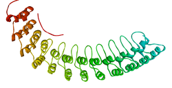
 Ribbon diagram of a fragment of the membrane-binding domain of ankyrin R.[1] | |||||||
| Identifiers | |||||||
| Symbol | ANK1 | ||||||
| Alt. symbols | AnkyrinR, Band2.1 | ||||||
| NCBI gene | 286 | ||||||
| HGNC | 492 | ||||||
| OMIM | 182900 | ||||||
| PDB | 1N11 | ||||||
| RefSeq | NM_000037 | ||||||
| UniProt | P16157 | ||||||
| Other data | |||||||
| Locus | Chr. 8 p21.1-11.2 | ||||||
| |||||||
| Ankyrin repeat | |||||||||
|---|---|---|---|---|---|---|---|---|---|
| Identifiers | |||||||||
| Symbol | Ank | ||||||||
| Pfam | PF00023 | ||||||||
| InterPro | IPR002110 | ||||||||
| SMART | SM00248 | ||||||||
| PROSITE | PDOC50088 | ||||||||
| SCOP2 | 1awc / SCOPe / SUPFAM | ||||||||
| |||||||||
| ANK2, neuronal | |||||||
|---|---|---|---|---|---|---|---|
| Identifiers | |||||||
| Symbol | ANK2 | ||||||
| Alt. symbols | AnkyrinB | ||||||
| NCBI gene | 287 | ||||||
| HGNC | 493 | ||||||
| OMIM | 106410 | ||||||
| RefSeq | NM_001148 | ||||||
| UniProt | Q01484 | ||||||
| Other data | |||||||
| Locus | Chr. 4 q25-q27 | ||||||
| |||||||
| ANK3, node of Ranvier | |||||||
|---|---|---|---|---|---|---|---|
| Identifiers | |||||||
| Symbol | ANK3 | ||||||
| Alt. symbols | AnkyrinG | ||||||
| NCBI gene | 288 | ||||||
| HGNC | 494 | ||||||
| OMIM | 600465 | ||||||
| RefSeq | NM_020987 | ||||||
| UniProt | Q12955 | ||||||
| Other data | |||||||
| Locus | Chr. 10 q21 | ||||||
| |||||||
Ankyrins are a family of proteins that mediate the attachment of integral membrane proteins to the spectrin-actin based membrane cytoskeleton.[2] Ankyrins have binding sites for the beta subunit of spectrin and at least 12 families of integral membrane proteins. This linkage is required to maintain the integrity of the plasma membranes and to anchor specific ion channels, ion exchangers and ion transporters in the plasma membrane. The name is derived from the Greek word ἄγκυρα (ankyra) for "anchor".
- ^ PDB: 1N11; Michaely P, Tomchick DR, Machius M, Anderson RG (December 2002). "Crystal structure of a 12 ANK repeat stack from human ankyrinR". The EMBO Journal. 21 (23): 6387–96. doi:10.1093/emboj/cdf651. PMC 136955. PMID 12456646.
- ^ Bennett V, Baines AJ (July 2001). "Spectrin and ankyrin-based pathways: metazoan inventions for integrating cells into tissues". Physiological Reviews. 81 (3): 1353–92. doi:10.1152/physrev.2001.81.3.1353. PMID 11427698. S2CID 15307181.