| Cementoenamel junction | |
|---|---|
 Labeled molar | |
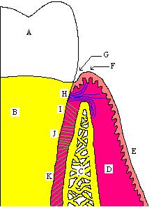
 The CEJ is the more or less horizontal demarcation line that distinguishes the crown (A) of the tooth from root (B) of the tooth. | |
| Identifiers | |
| MeSH | D019237 |
| TA2 | 926 |
| FMA | 55627 |
| Anatomical terminology | |
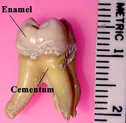
In dental anatomy, the cementoenamel junction (CEJ) is the location where the enamel, which covers the anatomical crown of a tooth, and the cementum, which covers the anatomical root of a tooth, meet. Informally it is known as the neck of the tooth.[1] The border created by these two dental tissues has much significance as it is usually the location where the gingiva (gums) attaches to a healthy tooth by fibers called the gingival fibers.[2]
Active recession of the gingiva reveals the cementoenamel junction in the mouth and is usually a sign of an unhealthy condition. The loss of attachment is considered a more reliable indicator of periodontal disease. The CEJ is the site of major tooth resorption. A significant proportion of tooth loss is caused by tooth resorption, which occurs in 5 to 10 percent of the population. The clinical location of CEJ which is a static landmark, serves as a crucial anatomical site for the measurement of probing pocket depth (PPD) and clinical attachment level (CAL). The CEJ varies between subjects, but also between teeth from the same person.[1]
There exists a normal variation in the relationship of the cementum and the enamel at the cementoenamel junction. In about 60–65% of teeth, the cementum overlaps the enamel at the CEJ, while in about 30% of teeth, the cementum and enamel abut each other with no overlap. In only 5–10% of teeth, there is a space between the enamel and the cementum at which the underlying dentin is exposed.[3]
- ^ a b Vandana KL, Haneet RK (September 2014). "Cementoenamel junction: An insight". Journal of Indian Society of Periodontology. 18 (5): 549–554. doi:10.4103/0972-124X.142437. PMC 4239741. PMID 25425813.
- ^ Clemente CD (1987). Anatomy, a regional atlas of the human body. Internet Archive. Baltimore : Urban & Schwarzenberg. ISBN 978-0-8067-0323-7.
- ^ Carranza FA, Bernard GW (2002). "The Tooth-Supporting Structures". In Newman MG, Takei HH, Carranza FA (eds.). Carranza's Clinical Periodontology (9th ed.). Philadelphia: W. B. Saunders. p. 43. ISBN 978-0-7216-8331-7.