| Cell biology | |
|---|---|
| centrosome | |
 Components of a typical centrosome:
|

In cell biology a centriole is a cylindrical organelle composed mainly of a protein called tubulin.[1] Centrioles are found in most eukaryotic cells, but are not present in conifers (Pinophyta), flowering plants (angiosperms) and most fungi, and are only present in the male gametes of charophytes, bryophytes, seedless vascular plants, cycads, and Ginkgo.[2][3] A bound pair of centrioles, surrounded by a highly ordered mass of dense material, called the pericentriolar material (PCM),[4] makes up a structure called a centrosome.[1]
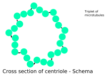
Centrioles are typically made up of nine sets of short microtubule triplets, arranged in a cylinder. Deviations from this structure include crabs and Drosophila melanogaster embryos, with nine doublets, and Caenorhabditis elegans sperm cells and early embryos, with nine singlets.[5][6] Additional proteins include centrin, cenexin and tektin.[7]
The main function of centrioles is to produce cilia during interphase and the aster and the spindle during cell division.
- ^ a b Eddé, B; Rossier, J; Le Caer, JP; Desbruyères, E; Gros, F; Denoulet, P (1990). "Posttranslational glutamylation of alpha-tubulin". Science. 247 (4938): 83–5. Bibcode:1990Sci...247...83E. doi:10.1126/science.1967194. PMID 1967194.
- ^ Quarmby, LM; Parker, JD (2005). "Cilia and the cell cycle?". The Journal of Cell Biology. 169 (5): 707–10. doi:10.1083/jcb.200503053. PMC 2171619. PMID 15928206.
- ^ Silflow, CD; Lefebvre, PA (2001). "Assembly and motility of eukaryotic cilia and flagella. Lessons from Chlamydomonas reinhardtii". Plant Physiology. 127 (4): 1500–1507. doi:10.1104/pp.010807. PMC 1540183. PMID 11743094.
- ^ Lawo, Steffen; Hasegan, Monica; Gupta, Gagan D.; Pelletier, Laurence (November 2012). "Subdiffraction imaging of centrosomes reveals higher-order organizational features of pericentriolar material". Nature Cell Biology. 14 (11): 1148–1158. doi:10.1038/ncb2591. ISSN 1476-4679. PMID 23086237. S2CID 11286303.
- ^ Delattre, M; Gönczy, P (2004). "The arithmetic of centrosome biogenesis" (PDF). Journal of Cell Science. 117 (Pt 9): 1619–30. doi:10.1242/jcs.01128. PMID 15075224. S2CID 7046196. Archived (PDF) from the original on 18 August 2017.
- ^ Leidel, S.; Delattre, M.; Cerutti, L.; Baumer, K.; Gönczy, P (2005). "SAS-6 defines a protein family required for centrosome duplication in C. elegans and in human cells". Nature Cell Biology. 7 (2): 115–25. doi:10.1038/ncb1220. PMID 15665853. S2CID 4634352.
- ^ Rieder, C. L.; Faruki, S.; Khodjakov, A. (October 2001). "The centrosome in vertebrates: more than a microtubule-organizing center". Trends in Cell Biology. 11 (10): 413–419. doi:10.1016/S0962-8924(01)02085-2. ISSN 0962-8924. PMID 11567874.