| Choroid | |
|---|---|
 Cross-section of human eye, with choroid labeled at top. | |
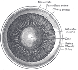
 Interior of anterior half of bulb of eye. (Choroid labeled at right, second from the bottom.) | |
| Details | |
| Artery | Short posterior ciliary arteries, long posterior ciliary arteries |
| Identifiers | |
| Latin | choroidea |
| MeSH | D002829 |
| TA98 | A15.2.03.002 |
| TA2 | 6774 |
| FMA | 58298 |
| Anatomical terminology | |
The choroid, also known as the choroidea or choroid coat, is a part of the uvea, the vascular layer of the eye. It contains connective tissues, and lies between the retina and the sclera. The human choroid is thickest at the far extreme rear of the eye (at 0.2 mm), while in the outlying areas it narrows to 0.1 mm.[1] The choroid provides oxygen and nourishment to the outer layers of the retina. Along with the ciliary body and iris, the choroid forms the uveal tract.
The structure of the choroid is generally divided into four layers (classified in order of furthest away from the retina to closest):
- Haller's layer – outermost layer of the choroid consisting of larger diameter blood vessels;[1]
- Sattler's layer – layer of medium diameter blood vessels;[1]
- Choriocapillaris – layer of capillaries;[1] and
- Bruch's membrane (synonyms: Lamina basalis, Complexus basalis, Lamina vitra) – innermost layer of the choroid.[1]