| Clubfoot | |
|---|---|
| Other names | Clubfeet, congenital talipes equinovarus (CTEV)[1] |
 | |
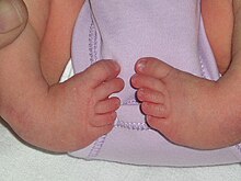
| Bilateral clubfeet | |
| Specialty | Orthopedics, podiatry |
| Symptoms | Foot that is rotated inwards and downwards[2] |
| Usual onset | During early pregnancy[1] |
| Causes | Unknown[1] |
| Risk factors | Genetics, mothers who smoke cigarettes, males,[1] ethnicity |
| Diagnostic method | Physical examination, ultrasound during pregnancy[1][3] |
| Differential diagnosis | Metatarsus adductus[4] |
| Treatment | Ponseti method (manipulation, casting, cutting the Achilles tendon, braces), French method, surgery[1][3] |
| Prognosis | Good with proper treatment[3] |
| Frequency | 1 in 1,000[3] |
Clubfoot is a congenital or acquired defect where one or both feet are rotated inward and downward.[1][2] Congenital clubfoot is the most common congenital malformation of the foot with an incidence of 1 per 1000 births.[5] In approximately 50% of cases, clubfoot affects both feet, but it can present unilaterally causing one leg or foot to be shorter than the other.[1][6] Most of the time, it is not associated with other problems.[1] Without appropriate treatment, the foot deformity will persist and lead to pain and impaired ability to walk, which can have a dramatic impact on the quality of life.[5][3][7]
The exact cause is usually not identified.[1][3] Both genetic and environmental factors are believed to be involved.[1][3] There are two main types of congenital clubfoot: idiopathic (80% of cases) and secondary clubfoot (20% of cases). The idiopathic congenital clubfoot is a multifactorial condition that includes environmental, vascular, positional, and genetic factors.[8] There appears to be hereditary component for this birth defect given that the risk of developing congenital clubfoot is 25% when a first-degree relative is affected.[8] In addition, if one identical twin is affected, there is a 33% chance the other one will be as well.[1] The underlying mechanism involves disruption of the muscles or connective tissue of the lower leg, leading to joint contracture.[1][9] Other abnormalities are associated 20% of the time, with the most common being distal arthrogryposis and myelomeningocele.[1][3] The diagnosis may be made at birth by physical examination or before birth during an ultrasound exam.[1][3]
The most common initial treatment is the Ponseti method, which is divided into two phases: 1) correcting of foot position and 2) casting at repeated weekly intervals.[1] If the clubfoot deformity does not improve by the end of the casting phase, an Achilles tendon tenotomy can be performed.[10] The procedure consists of a small posterior skin incision through which the tendon cut is made. In order to maintain the correct position of the foot, it is necessary to wear an orthopedic brace until 5 years of age.[11]
Initially, the brace is worn nearly continuously and then just at night.[1] In about 20% of cases, further surgery is required.[1] Treatment can be carried out by a range of healthcare providers and can generally be achieved in the developing world with few resources.[1]
Congenital clubfoot occurs in 1 to 4 of every 1,000 live births, making it one of the most common birth defects affecting the legs.[6][3][7] About 80% of cases occur in developing countries where there is limited access to care.[6] Clubfoot is more common in firstborn children and males.[1][6][7] It is more common among Māori people, and less common among Chinese people.[3]
- ^ a b c d e f g h i j k l m n o p q r s t Gibbons PJ, Gray K (September 2013). "Update on clubfoot". Journal of Paediatrics and Child Health. 49 (9): E434–E437. doi:10.1111/jpc.12167. PMID 23586398. S2CID 6185031.
- ^ a b "Talipes equinovarus". Genetic and Rare Diseases Information Center (GARD). 2017. Archived from the original on October 15, 2017. Retrieved October 15, 2017.
- ^ a b c d e f g h i j k Dobbs MB, Gurnett CA (May 2009). "Update on clubfoot: etiology and treatment". Clinical Orthopaedics and Related Research. 467 (5): 1146–1153. doi:10.1007/s11999-009-0734-9. PMC 2664438. PMID 19224303.
- ^ Moses S. "Clubfoot". www.fpnotebook.com. Archived from the original on October 15, 2017. Retrieved October 15, 2017.
- ^ a b Dibello D, Di Carlo V, Colin G, Barbi E, Galimberti AM (June 2020). "What a paediatrician should know about congenital clubfoot". Italian Journal of Pediatrics. 46 (1): 78. doi:10.1186/s13052-020-00842-3. PMC 7271518. PMID 32498693.
- ^ a b c d Smythe T, Kuper H, Macleod D, Foster A, Lavy C (March 2017). "Birth prevalence of congenital talipes equinovarus in low- and middle-income countries: a systematic review and meta-analysis". Tropical Medicine & International Health. 22 (3): 269–285. doi:10.1111/tmi.12833. PMID 28000394.
- ^ a b c O'Shea RM, Sabatini CS (December 2016). "What is new in idiopathic clubfoot?". Current Reviews in Musculoskeletal Medicine. 9 (4): 470–477. doi:10.1007/s12178-016-9375-2. PMC 5127955. PMID 27696325.
- ^ a b Basit S, Khoshhal KI (February 2018). "Genetics of clubfoot; recent progress and future perspectives". European Journal of Medical Genetics. 61 (2): 107–113. doi:10.1016/j.ejmg.2017.09.006. PMID 28919208.
- ^ Cummings RJ, Davidson RS, Armstrong PF, Lehman WB (February 2002). "Congenital clubfoot". The Journal of Bone and Joint Surgery. American Volume. 84 (2): 290–308. doi:10.2106/00004623-200202000-00018. PMID 11861737.
- ^ Ganesan B, Luximon A, Al-Jumaily A, Balasankar SK, Naik GR (June 20, 2017). Nazarian A (ed.). "Ponseti method in the management of clubfoot under 2 years of age: A systematic review". PLOS ONE. 12 (6): e0178299. Bibcode:2017PLoSO..1278299G. doi:10.1371/journal.pone.0178299. PMC 5478104. PMID 28632733.
- ^ Morcuende JA, Abbasi D, Dolan LA, Ponseti IV (September 2005). "Results of an accelerated Ponseti protocol for clubfoot". Journal of Pediatric Orthopedics. 25 (5): 623–626. doi:10.1097/01.bpo.0000162015.44865.5e. PMID 16199943. S2CID 25067281.