| Corneal | |
|---|---|
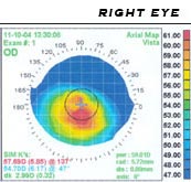
 A corneal topogram of an eye affected by keratoconus. Blue shows the flattest areas, and red the steepest. | |
| MeSH | D019781 |
Corneal topography, also known as photokeratoscopy or videokeratography, is a non-invasive medical imaging technique for mapping the anterior curvature of the cornea, the outer structure of the eye. Since the cornea is normally responsible for some 70% of the eye's refractive power,[1] its topography is of critical importance in determining the quality of vision and corneal health.
The three-dimensional map is therefore a valuable aid to the examining ophthalmologist or optometrist and can assist in the diagnosis and treatment of a number of conditions; in planning cataract surgery and intraocular lens implantation; in planning refractive surgery such as LASIK, and evaluating its results; or in assessing the fit of contact lenses. A development of keratoscopy, corneal topography extends the measurement range from the four points a few millimeters apart that is offered by keratometry to a grid of thousands of points covering the entire cornea. The procedure is carried out in seconds and is painless.
- ^ Pavan-Langston, Deborah (2007). Manual of Ocular Diagnosis and Therapy. Hagerstown, MD: Lippincott Williams & Wilkins. p. 405. ISBN 978-0-7817-6512-1.