| Cutaneous myxoma | |
|---|---|
| Other names | Superficial angiomyxoma |
 | |
| Cutaneous myxoma | |
| Specialty | Oncology |
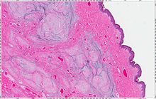
A cutaneous myxoma, or superficial angiomyxoma, consists of a multilobulated myxoid mass containing stellate or spindled fibroblasts with pools of mucin forming cleft-like spaces. There is often a proliferation of blood vessels and an inflammatory infiltrate. Staining is positive for vimentin, negative for cytokeratin and desmin, and variable for CD34, Factor VIIIa, SMA, MSA and S-100.[1]
Clinically, it may present as solitary or multiple flesh-colored nodules on the face, trunk, or extremities. It may occur as part of the Carney complex, and is sometimes the first sign. Local recurrence is common.[2] Cutaneous myxoma is diagnosed based on histopathological features. The differential diagnosis for cutaneous myxoma include alopecia areata, verrucous hamartoma, cyst, fibroma, glioma, hemangioma, lipoma, scar, and nevus sebaceous. Treatment involves complete surgical excision.
- ^ Satter EK (October 2009). "Solitary superficial angiomyxoma: an infrequent but distinct soft tissue tumor". J. Cutan. Pathol. 36 (Suppl 1): 56–9. doi:10.1111/j.1600-0560.2008.01216.x. PMID 19187115. S2CID 1528140. Archived from the original on 2012-12-10.
- ^ James, William; Berger, Timothy; Elston, Dirk (2005). Andrews' Diseases of the Skin: Clinical Dermatology (10th ed.). Saunders. p. 614. ISBN 0-7216-2921-0.