| Ductus venosus | |
|---|---|
 | |
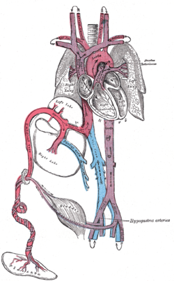
 The liver and the veins in connection with it, of a human embryo, twenty-four or twenty-five days old, as seen from the ventral surface. | |
| Details | |
| Source | Umbilical vein |
| Drains to | Inferior vena cava |
| Artery | Ductus arteriosus |
| Identifiers | |
| Latin | ductus venosus |
| Anatomical terminology | |
In the fetus, the ductus venosus (Arantius' duct after Julius Caesar Aranzi[1]) shunts a portion of umbilical vein blood flow directly to the inferior vena cava.[2] Thus, it allows oxygenated blood from the placenta to bypass the liver. Compared to the 50% shunting of umbilical blood through the ductus venosus found in animal experiments, the degree of shunting in the human fetus under physiological conditions is considerably less, 30% at 20 weeks, which decreases to 18% at 32 weeks, suggesting a higher priority of the fetal liver than previously realized.[3] In conjunction with the other fetal shunts, the foramen ovale and ductus arteriosus, it plays a critical role in preferentially shunting oxygenated blood to the fetal brain. It is a part of fetal circulation.
- ^ "Whonamedit - dictionary of medical eponyms". www.whonamedit.com.
- ^ Kiserud, T.; Rasmussen, S.; Skulstad, S. (2000). "Blood flow and the degree of shunting through the ductus venosus in the human fetus". American Journal of Obstetrics and Gynecology. 182 (1 Pt 1): 147–153. doi:10.1016/S0002-9378(00)70504-7. PMID 10649170.
- ^ Kiserud, T (2000). "Fetal venous circulation -- an update on hemodynamics". J Perinat Med. 28 (2): 90–6. doi:10.1515/JPM.2000.011. PMID 10875092. S2CID 11799576.