| Cell biology | |
|---|---|
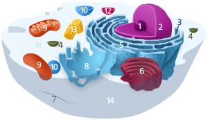
| Animal cell diagram | |
 Components of a typical animal cell:
|

The endoplasmic reticulum (ER) is a part of a transportation system of the eukaryotic cell, and has many other important functions such as protein folding. It is a type of organelle made up of two subunits – rough endoplasmic reticulum (RER), and smooth endoplasmic reticulum (SER). The endoplasmic reticulum is found in most eukaryotic cells and forms an interconnected network of flattened, membrane-enclosed sacs known as cisternae (in the RER), and tubular structures in the SER. The membranes of the ER are continuous with the outer nuclear membrane. The endoplasmic reticulum is not found in red blood cells, or spermatozoa.
The two types of ER share many of the same proteins and engage in certain common activities such as the synthesis of certain lipids and cholesterol. Different types of cells contain different ratios of the two types of ER depending on the activities of the cell. RER is found mainly toward the nucleus of cell and SER towards the cell membrane or plasma membrane of cell.
The outer (cytosolic) face of the RER is studded with ribosomes that are the sites of protein synthesis. The RER is especially prominent in cells such as hepatocytes. The SER lacks ribosomes and functions in lipid synthesis but not metabolism, the production of steroid hormones, and detoxification.[1] The SER is especially abundant in mammalian liver and gonad cells.
The ER was observed by light microscopy by Garnier in 1897, who coined the term ergastoplasm.[2][3] The lacy membranes of the endoplasmic reticulum were first seen by electron microscopy in 1945 by Keith R. Porter, Albert Claude, and Ernest F. Fullam.[4] Later, the word reticulum, which means "network", was applied by Porter in 1953 to describe this fabric of membranes.[5]
- ^ "Endoplasmic Reticulum (Rough and Smooth)". British Society of Cell Biology. Archived from the original on 24 November 2015. Retrieved 21 November 2015.
- ^ Garnier, C. (1897). "Les filaments basaux des cellules glandulaires. Note préliminaire". Bibliographie Anatomique. 5: 278–289. OCLC 493441682.
- ^ Buvat, R. (1963). "Electron Microscopy of Plant Protoplasm". International Review of Cytology Volume 14. Vol. 14. pp. 41–155. doi:10.1016/S0074-7696(08)60021-2. ISBN 978-0-12-364314-8. PMID 14283576.
- ^ Porter KR, Claude A, Fullam EF (March 1945). "A study of tissue culture cells by electron microscopy: methods and preliminary observations". The Journal of Experimental Medicine. 81 (3): 233–46. doi:10.1084/jem.81.3.233. PMC 2135493. PMID 19871454.
- ^ PORTER KR (May 1953). "Observations on a submicroscopic basophilic component of cytoplasm". The Journal of Experimental Medicine. 97 (5): 727–50. doi:10.1084/jem.97.5.727. PMC 2136295. PMID 13052830.