| Functional magnetic resonance imaging | |
|---|---|
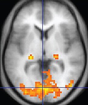
 An fMRI image with yellow areas showing increased activity compared with a control condition | |
| Purpose | Measures brain activity detecting changes due to blood flow. |
Functional magnetic resonance imaging or functional MRI (fMRI) measures brain activity by detecting changes associated with blood flow.[1][2] This technique relies on the fact that cerebral blood flow and neuronal activation are coupled. When an area of the brain is in use, blood flow to that region also increases.[3]
The primary form of fMRI uses the blood-oxygen-level dependent (BOLD) contrast,[4] discovered by Seiji Ogawa in 1990. This is a type of specialized brain and body scan used to map neural activity in the brain or spinal cord of humans or other animals by imaging the change in blood flow (hemodynamic response) related to energy use by brain cells.[4] Since the early 1990s, fMRI has come to dominate brain mapping research because it does not involve the use of injections, surgery, the ingestion of substances, or exposure to ionizing radiation.[5] This measure is frequently corrupted by noise from various sources; hence, statistical procedures are used to extract the underlying signal. The resulting brain activation can be graphically represented by color-coding the strength of activation across the brain or the specific region studied. The technique can localize activity to within millimeters but, using standard techniques, no better than within a window of a few seconds.[6] Other methods of obtaining contrast are arterial spin labeling[7] and diffusion MRI. Diffusion MRI is similar to BOLD fMRI but provides contrast based on the magnitude of diffusion of water molecules in the brain.
In addition to detecting BOLD responses from activity due to tasks or stimuli, fMRI can measure resting state, or negative-task state, which shows the subjects' baseline BOLD variance. Since about 1998 studies have shown the existence and properties of the default mode network, a functionally connected neural network of apparent resting brain states.
fMRI is used in research, and to a lesser extent, in clinical work. It can complement other measures of brain physiology such as electroencephalography (EEG), and near-infrared spectroscopy (NIRS). Newer methods which improve both spatial and time resolution are being researched, and these largely use biomarkers other than the BOLD signal. Some companies have developed commercial products such as lie detectors based on fMRI techniques, but the research is not believed to be developed enough for widespread commercial use.[8]
- ^ "Magnetic Resonance, a critical peer-reviewed introduction; functional MRI" (PDF). TRTF/EMRF 2023. Retrieved 23 January 2023.
- ^ Huettel, Song & McCarthy (2009)
- ^ Logothetis, N. K.; Pauls, Jon; Augath, M.; Trinath, T.; Oeltermann, A. (July 2001). "A neurophysiological investigation of the basis of the BOLD signal in fMRI". Nature. 412 (6843): 150–157. Bibcode:2001Natur.412..150L. doi:10.1038/35084005. PMID 11449264. S2CID 969175.
Our results show unequivocally that a spatially localized increase in the BOLD contrast directly and monotonically reflects an increase in neural activity.
- ^ a b Huettel, Song & McCarthy (2009, p. 26)
- ^ Huettel, Song & McCarthy (2009, p. 4)
- ^ Thomas, Roger K (1 January 1993). "INTRODUCTION: A Biopsychology Festschrift in Honor of Lelon J. Peacock". Journal of General Psychology. 120 (1): 5.
- ^ Detre, John A.; Rao, Hengyi; Wang, Danny J.J.; Chen, Yu Fen; Wang, Ze (May 2012). "Applications of arterial spin labeled MRI in the brain". Journal of Magnetic Resonance Imaging. 35 (5): 1026–1037. doi:10.1002/jmri.23581. PMC 3326188. PMID 22246782.
- ^ Langleben, D. D.; Moriarty, J. C. (2013). "Using Brain Imaging for Lie Detection: Where Science, Law and Research Policy Collide". Psychol Public Policy Law. 19 (2): 222–234. doi:10.1037/a0028841. PMC 3680134. PMID 23772173.