| Infantile hemangioma | |
|---|---|
| Other names | Infantile haemangioma, hemangioma, capillary hemangioma, capillary angioma, strawberry haemangioma, strawberry mark [1][2] |
 | |
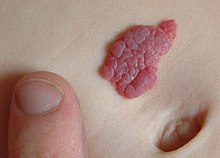
| A small hemangioma of infancy | |
| Specialty | Dermatology, gastroenterology, Oral and Maxillofacial Surgery |
| Symptoms | Raised red or blue lesion[3] |
| Complications | Pain, bleeding, ulcer formation, heart failure, disfigurement[1] |
| Usual onset | First 4 weeks of life[1] |
| Types | Superficial, deep, mixed[1] |
| Risk factors | Females, whites, premature birth, low birth weight babies[1] |
| Diagnostic method | Based on symptoms and appearance[1] |
| Differential diagnosis | Congenital hemangioma, pyogenic granuloma, kaposiform hemangioendothelioma, tufted angioma, venous malformation,[1] other vascular anomaly[4] |
| Treatment | Close observation, medication[5][1] |
| Medication | Propranolol, Timolol, steroids[5][1] |
| Frequency | Up to 5%[5] |
An infantile hemangioma (IH), sometimes called a strawberry mark due to appearance, is a type of benign vascular tumor or anomaly that affects babies.[1][2] Other names include capillary hemangioma,[6] "strawberry hemangioma",[7]: 593 strawberry birthmark[8] and strawberry nevus.[6] and formerly known as a cavernous hemangioma. They appear as a red or blue raised lesion on the skin.[3] Typically, they begin during the first four weeks of life,[9] growing until about five months of life,[10] and then shrinking in size and disappearing over the next few years.[1][2] Often skin changes remain after they shrink.[1][5] Complications may include pain, bleeding, ulcer formation, disfigurement, or heart failure.[1] It is the most common tumor of orbit and periorbital areas in childhood. It may occur in the skin, subcutaneous tissues and mucous membranes of oral cavities and lips as well as in extracutaneous locations including the liver and gastrointestinal tract.
The underlying reason for their occurrence is not clear.[1] In about 10% of cases they appear to run in families.[1] A few cases are associated with other abnormalities such as PHACE syndrome.[1] Diagnosis is generally based on the symptoms and appearance.[1] Occasionally medical imaging can assist in the diagnosis.[1]
In most cases no treatment is needed, other than close observation.[5][1] Hemangiomas may grow rapidly, before stopping and slowly fading, with maximum improvement typically occurring by the age of 3.5 years.[11][12] While this birthmark may be alarming in appearance, physicians generally counsel that it be left to disappear on its own, unless it is in the way of vision or blocking the nostrils.[9] Certain cases, however, may result in problems and the use of medication such as propranolol or steroids are recommended.[5][1] Occasionally surgery or laser treatment may be used.[1]
It is one of the most common benign tumors in babies, occurring in about 5-10% of all births.[5][1][13]: 81 They occur more frequently in females, whites,[14][15] prematurely born,[14][15] and low birth weight babies.[5][1] They can occur anywhere on the body, though 83% occur on the head or neck area.[14] The word "hemangioma" comes from the Greek haima (αἷμα) meaning "blood"; angeion (ἀγγεῖον) meaning "vessel"; and -oma (-ωμα) meaning "tumor".[16]
- ^ a b c d e f g h i j k l m n o p q r s t u v w Darrow DH, Greene AK, Mancini AJ, Nopper AJ (October 2015). "Diagnosis and Management of Infantile Hemangioma". Pediatrics. 136 (4): e1060-104. doi:10.1542/peds.2015-2485. PMID 26416931.
- ^ a b c "Birthmarks NHS". nhs.uk. 2017-10-20. Retrieved 2021-04-16.
- ^ a b "Infantile Hemangiomas". Merck Manuals Professional Edition. Retrieved 7 January 2019.
- ^ Workshop I. "ISSVA Classification of Vascular Anomalies" (PDF). ISSVA Classification pdf. ISSVA. Retrieved 23 March 2022.
- ^ a b c d e f g h Krowchuk DP, Frieden IJ, Mancini AJ, Darrow DH, Blei F, Greene AK, Annam A, Baker CN, Frommelt PC, Hodak A, Pate BM, Pelletier JL, Sandrock D, Weinberg ST, Whelan MA, SUBCOMMITTEE ON THE MANAGEMENT OF INFANTILE H (January 2019). "Clinical Practice Guideline for the Management of Infantile Hemangiomas". Pediatrics. 143 (1): e20183475. doi:10.1542/peds.2018-3475. PMID 30584062.
- ^ a b Ronald P R, Jean L B, Joseph L J (2007). Dermatology: 2-Volume Set. St. Louis: Mosby. ISBN 978-1-4160-2999-1.
- ^ James W, Berger T, Elston D (2005). Andrews' Diseases of the Skin: Clinical Dermatology (10th ed.). Saunders. ISBN 0-7216-2921-0.
- ^ "What is Infantile Hemangioma (Strawberry Birthmark)?".
- ^ a b "Birthmarks". Parenting and Child Health website. Archived from the original on 2008-07-23. Retrieved 2008-08-02.
- ^ Park M (2021). "Infantile hemangioma: Timely diagnosis and treatment". Clinical and Experimental Pediatrics. 64 (11): 573–574. doi:10.3345/cep.2021.00752. PMC 8566800. PMID 34325500.
- ^ Cite error: The named reference
Couto2012was invoked but never defined (see the help page). - ^ Wahrman JE (1994). "Hemangiomas". Pediatr Rev. 15 (7): 266–271. doi:10.1542/pir.15-7-266. PMID 8084847.
- ^ Sadler TW (2009). Langman's Medical Embryology (11th ed.). Lippincott Williams & Wilkins. ISBN 978-1-60547-656-8.
- ^ a b c "Hemangioma Information". Vascular Birthmark Foundation. Retrieved 2008-08-02.
- ^ a b "Birthmarks". American Academy of Dermatology. Retrieved 2008-08-02.
- ^ Lexicon Orthopaedic Etymology. CRC Press. 1999. p. 16. ISBN 978-90-5702-597-6.