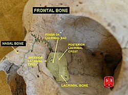
| Lacrimal bone | |
|---|---|
 Position of the lacrimal bone (shown in green). | |
 Medial wall of the orbit. Lacrimal bone is in yellow. | |
| Details | |
| Identifiers | |
| Latin | os lacrimale |
| TA98 | A02.1.09.001 |
| TA2 | 744 |
| FMA | 52741 |
| Anatomical terms of bone | |
The lacrimal bones are two small and fragile bones of the facial skeleton; they are roughly the size of the little fingernail and situated at the front part of the medial wall of the orbit. They each have two surfaces and four borders. Several bony landmarks of the lacrimal bones function in the process of lacrimation. Specifically, the lacrimal bones help form the nasolacrimal canal necessary for tear translocation. A depression on the anterior inferior portion of one bone, the lacrimal fossa, houses the membranous lacrimal sac. Tears, from the lacrimal glands, collect in this sac during excessive lacrimation. The fluid then flows through the nasolacrimal duct and into the nasopharynx. This drainage results in what is commonly referred to a runny nose during excessive crying or tear production. Injury or fracture of the lacrimal bone can result in posttraumatic obstruction of the lacrimal pathways.[1][2]
- ^ Maliborski, A; Różycki, R (2014). "Diagnostic imaging of the nasolacrimal drainage system. Part I. Radiological anatomy of lacrimal pathways. Physiology of tear secretion and tear outflow". Med. Sci. Monit. 20: 628–38. doi:10.12659/MSM.890098. PMC 3999077. PMID 24743297.
- ^ Saladin (7 January 2014). Anatomy & Physiology : The Unity of Form and Function (Seventh ed.). New York: McGraw-Hill Education. ISBN 978-0-07-340371-7.[page needed]