| Eye | |
|---|---|
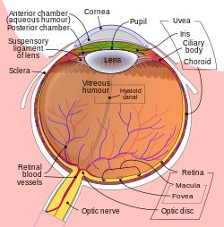
 Schematic diagram of the human eye. | |
 | |
| Details | |
| Identifiers | |
| Latin | oculus (plural: oculi) |
| Anatomical terminology | |

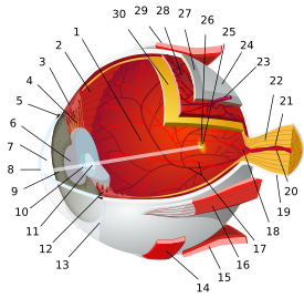
- posterior segment
- ora serrata
- ciliary muscle
- ciliary zonules
- Schlemm's canal
- pupil
- anterior chamber
- cornea
- iris
- lens cortex
- lens nucleus
- ciliary process
- conjunctiva
- inferior oblique muscle
- inferior rectus muscle
- medial rectus muscle
- retinal arteries and veins
- optic disc
- dura mater
- central retinal artery
- central retinal vein
- optic nerve
- vorticose vein
- bulbar sheath
- macula
- fovea
- sclera
- choroid
- superior rectus muscle
- retina
Mammals normally have a pair of eyes. Although mammalian vision is not as excellent as bird vision, it is at least dichromatic for most of mammalian species, with certain families (such as Hominidae) possessing a trichromatic color perception.
The dimensions of the eyeball vary only 1–2 mm among humans. The vertical axis is 24 mm, while the transverse is larger. At birth it is generally 16–17 mm, enlarging to 22.5–23 mm by three years of age. Between birth and age 13 the eye attains its mature size. It weighs 7.5 grams and its volume is roughly 6.5 ml. Along a line through the nodal (central) point of the eye is the optic axis, which is at a slight slant of five degrees toward the nose from the visual axis (i.e., the section going towards the focused point of the fovea).