Marcello Malpighi | |
|---|---|
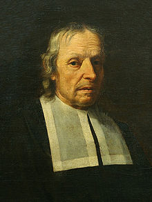
 Marcello Malpighi, a portrait from life by Carlo Cignani | |
| Born | 10 March 1628 |
| Died | 30 November 1694 (aged 66) |
| Nationality | Italian |
| Alma mater | University of Bologna |
| Scientific career | |
| Fields | Anatomy, histology, physiology, embryology, medicine |
| Institutions | University of Bologna University of Pisa University of Messina |
| Doctoral advisor | Giovanni Alfonso Borelli |
| Doctoral students | Antonio Maria Valsalva |
Marcello Malpighi (10 March 1628 – 30 November 1694) was an Italian biologist and physician, who is referred to as the "Founder of microscopical anatomy, histology & Father of physiology and embryology". Malpighi's name is borne by several physiological features related to the biological excretory system, such as the Malpighian corpuscles and Malpighian pyramids of the kidneys and the Malpighian tubule system of insects. The splenic lymphoid nodules are often called the "Malpighian bodies of the spleen" or Malpighian corpuscles. The botanical family Malpighiaceae is also named after him. He was the first person to see capillaries in animals, and he discovered the link between arteries and veins that had eluded William Harvey. Malpighi was one of the earliest people to observe red blood cells under a microscope, after Jan Swammerdam. His treatise De polypo cordis (1666) was important for understanding blood composition, as well as how blood clots.[1] In it, Malpighi described how the form of a blood clot differed in the right against the left sides of the heart.[2]
The use of the microscope enabled Malpighi to discover that insects do not use lungs to breathe, but small holes in their skin called tracheae.[3] Malpighi also studied the anatomy of the brain and concluded this organ is a gland. In terms of modern endocrinology, this deduction is correct because the hypothalamus of the brain has long been recognized for its hormone-secreting capacity.[4]
Because Malpighi had a wide knowledge of both plants and animals, he made contributions to the scientific study of both. The Royal Society of London published two volumes of his botanical and zoological works in 1675 and 1679. Another edition followed in 1687, and a supplementary volume in 1697. In his autobiography, Malpighi speaks of his Anatome Plantarum, decorated with the engravings of Robert White, as "the most elegant format in the whole literate world."[5]
His study of plants led him to conclude that plants had tubules similar to those he saw in insects like the silkworm (using his microscope, he probably saw the stomata, through which plants exchange carbon dioxide with oxygen). Malpighi observed that when a ring-like portion of bark was removed on a trunk a swelling occurred in the tissues above the ring, and he correctly interpreted this as growth stimulated by food coming down from the leaves, and being blocked above the ring.[6]
- ^ Malpighi's De polypo cordis dissertatio (Treatise on cardiac polyp) was included as a chapter of his De viscerum structura exercitatio anatomica (Essay on the anatomical structure of the viscera, 1666).
- Malpighi, Marcello (1666). De Viscerum Structura Exercitatio Anatomica [Essay on the anatomical structure of the viscera] (in Latin). Bologna, (Italy): Giacomo Monti. pp. 151–172.
- English translation: Forrester, John M. (October 1995). "Malpighi's De polypo cordis: an annotated translation". Medical History. 39 (4): 477–492. doi:10.1017/s0025727300060385. PMC 1037031. PMID 8558994.
- ^ Lorraine Daston (2011). Histories of Scientific Observation. Chicago, USA: University of Chicago Press. p. 440. ISBN 978-0226136783.
- ^ Benjamin A. Rifkin and Michael J. Ackerman (2011). Human Anatomy: A Visual History from the Renaissance to the Digital Age. NY, USA: Abrams Books. p. 343. ISBN 978-0810997981.
- ^ Garabed Eknoyan, Natale Gaspare De Santo (1997). History of Nephrology 2: Reports from the First Congress on the International Association for the History of Nephrology, Kos, October 1996. Basel, Sewitzerland: S. Karger Publishing. p. 198. ISBN 978-3805564991.
- ^ Arber, Agnes (1942). "Nehemiah Grew (1641–1712) and Marcello Malpighi (1628–1694): an essay in comparison". Isis. 34 (1): 7–16. doi:10.1086/347742. JSTOR 225992. S2CID 143008947.
- ^ Domenico Bertoloni Meli (2011). Mechanism, Experiment, Disease: Marcello Malpighi and Seventeenth-Century Anatomy. Baltimore, USA: Johns Hopkins University Press. p. 456. ISBN 978-0801899041.