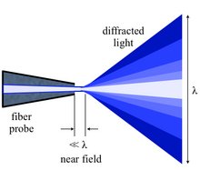

Near-field scanning optical microscopy (NSOM) or scanning near-field optical microscopy (SNOM) is a microscopy technique for nanostructure investigation that breaks the far field resolution limit by exploiting the properties of evanescent waves. In SNOM, the excitation laser light is focused through an aperture with a diameter smaller than the excitation wavelength, resulting in an evanescent field (or near-field) on the far side of the aperture.[3] When the sample is scanned at a small distance below the aperture, the optical resolution of transmitted or reflected light is limited only by the diameter of the aperture. In particular, lateral resolution of 6 nm[4] and vertical resolution of 2–5 nm have been demonstrated.[5][6]
As in optical microscopy, the contrast mechanism can be easily adapted to study different properties, such as refractive index, chemical structure and local stress. Dynamic properties can also be studied at a sub-wavelength scale using this technique.
NSOM/SNOM is a form of scanning probe microscopy.
- ^ Herzog JB (2011). Optical Spectroscopy of Colloidal CdSe Semiconductor Nanostructures (PDF) (Ph.D. thesis). University of Notre Dame.
- ^ Bao W, Borys NJ, Ko C, Suh J, Fan W, Thron A, et al. (August 2015). "Visualizing nanoscale excitonic relaxation properties of disordered edges and grain boundaries in monolayer molybdenum disulfide". Nature Communications. 6: 7993. Bibcode:2015NatCo...6.7993B. doi:10.1038/ncomms8993. PMC 4557266. PMID 26269394.
- ^ "SNOM || WITec". WITec Wissenschaftliche Instrumente und Technologie GmbH. Ulm Germany. Retrieved 2017-04-06.
- ^ Ma X, Liu Q, Yu N, Xu D, Kim S, Liu Z, et al. (November 2021). "6 nm super-resolution optical transmission and scattering spectroscopic imaging of carbon nanotubes using a nanometer-scale white light source". Nature Communications. 12 (1): 6868. arXiv:2006.04903. Bibcode:2021NatCo..12.6868M. doi:10.1038/s41467-021-27216-5. PMC 8617169. PMID 34824270.
- ^ Dürig U, Pohl DW, Rohner F (1986). "Near-field optical scanning microscopy". Journal of Applied Physics. 59 (10): 3318. Bibcode:1986JAP....59.3318D. doi:10.1063/1.336848.
- ^ Oshikane Y, Kataoka T, Okuda M, Hara S, Inoue H, Nakano M (April 2007). "Observation of nanostructure by scanning near-field optical microscope with small sphere probe" (free access). Science and Technology of Advanced Materials. 8 (3): 181. Bibcode:2007STAdM...8..181O. doi:10.1016/j.stam.2007.02.013.