| Pars intermedia | |
|---|---|
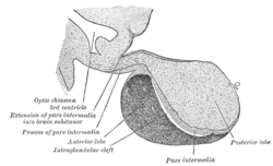
 Median sagittal through the hypophysis of an adult monkey. (Pars intermedia labeled at bottom center.) | |
| Details | |
| Identifiers | |
| Latin | pars intermedia adenohypophysis |
| TA98 | A11.1.00.004 A09.4.02.017 |
| TA2 | 3858 |
| TH | H3.08.02.2.00007 |
| FMA | 74632 |
| Anatomical terminology | |

The pars intermedia is one of the three parts of the anterior pituitary. It is a section of tissue sometimes called a middle or intermediate lobe, between the pars distalis, and the posterior pituitary.[1] It is a small region that is largely without blood supply.[2] The cells in the pars intermedia are large and pale. They surround follicles that contain a colloidal matrix.[3]
The pars intermedia secretes α-melanocyte-stimulating hormone (α-MSH), and corticotropin-like intermediate peptide.[citation needed] It appears to be tonically inhibited by the hypothalamus.
In the human fetus, this area produces melanocyte stimulating hormone (MSH) which causes the release of melanin produced in melanocytes that can give a darker skin pigmentation. In the adult the pars intermedia is either very small or entirely absent.
In less developed vertebrates the pars intermedia is much larger, and structurally and functionally more well defined.[4] In some animals including amphibians[5] it mediates active camouflage, causing darkening of the skin when placed against a darker background.
- ^ Ganapathy, MK; Tadi, P (January 2024). Anatomy, Head and Neck, Pituitary Gland. PMID 31855373.
- ^ Hall, John E.; Guyton, Arthur C. (2011). Guyton and Hall textbook of medical physiology (12th ed.). Philadelphia, Pa: Saunders/Elsevier. p. 895. ISBN 9781416045748.
- ^ Ilahi, Sadia; Ilahi, Tahir B. (3 October 2022). "Anatomy, Adenohypophysis (Pars Anterior, Anterior Pituitary)". Anatomy, Adenohypophysis (Pars Anterior, Anterior Pituitary). StatPearls Publishing. Retrieved 9 February 2023.
- ^ Hall, John E.; Guyton, Arthur C. (2011). Guyton and Hall textbook of medical physiology (12th ed.). Philadelphia, Pa: Saunders/Elsevier. p. 935. ISBN 9781416045748.
- ^ Davies, Andrew; Blakeley, Asa G. H.; Kidd, Cecil, eds. (2001). Human Physiology. pp. 397–398. ISBN 978-0-443-04559-2.