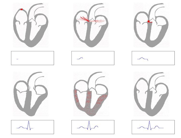
| Purkinje fibers | |
|---|---|
 Isolated heart conduction system showing Purkinje fibers | |
 The QRS complex is the large peak. | |
| Details | |
| Identifiers | |
| Latin | rami subendocardiales |
| MeSH | D011690 |
| TA98 | A12.1.06.008 |
| TA2 | 3961 |
| FMA | 9492 |
| Anatomical terminology | |
The Purkinje fibers, named for Jan Evangelista Purkyně, (English: /pɜːrˈkɪndʒi/ pur-KIN-jee;[1] Czech: [ˈpurkɪɲɛ] ; Purkinje tissue or subendocardial branches) are located in the inner ventricular walls of the heart,[2] just beneath the endocardium in a space called the subendocardium. The Purkinje fibers are specialized conducting fibers composed of electrically excitable cells.[3] They are larger than cardiomyocytes with fewer myofibrils and many mitochondria. They conduct cardiac action potentials more quickly and efficiently than any of the other cells in the heart's electrical conduction system.[4] Purkinje fibers allow the heart's conduction system to create synchronized contractions of its ventricles, and are essential for maintaining a consistent heart rhythm.[5]
- ^ Jones, Daniel (2011). Roach, Peter; Setter, Jane; Esling, John (eds.). Cambridge English Pronouncing Dictionary (18th ed.). Cambridge University Press. ISBN 978-0-521-15255-6.
- ^ Feher, Joseph (January 1, 2012), Feher, Joseph (ed.), "5.5 – The Cardiac Action Potential", Quantitative Human Physiology, Boston: Academic Press, pp. 458–466, doi:10.1016/b978-0-12-382163-8.00049-9, ISBN 978-0-12-382163-8, retrieved November 13, 2020
- ^ Stocum, David L. (January 1, 2012), Stocum, David L. (ed.), "Chapter 7 – Regeneration of Cardiac Muscle and Hematopoietic Tissues", Regenerative Biology and Medicine (Second Edition), San Diego: Academic Press, pp. 161–182, doi:10.1016/b978-0-12-384860-4.00007-1, ISBN 978-0-12-384860-4, retrieved November 13, 2020
- ^ "Purkinje fiber." The American Heritage® Medical Dictionary. 2007. Houghton Mifflin Company 23 Oct. 2016 http://medical-dictionary.thefreedictionary.com/Purkinje+fiber
- ^ Brooker, Graham (January 1, 2019), Segil, Jacob (ed.), "Chapter Fourteen – Pacemakers", Handbook of Biomechatronics, Academic Press, pp. 567–589, doi:10.1016/b978-0-12-812539-7.00014-3, ISBN 978-0-12-812539-7, S2CID 239396730, retrieved November 13, 2020