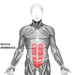
| Rectus abdominis | |
|---|---|
 The human rectus abdominis muscle. | |
| Details | |
| Origin | Crest of pubic |
| Insertion | Costal cartilages of ribs 5-7 xiphoid process of sternum |
| Artery | Inferior epigastric artery |
| Nerve | Segmentally by thoraco-abdominal nerves (T7 to T11) and subcostal (T12) |
| Actions | Flexion of the lumbar spine |
| Antagonist | Erector spinae |
| Identifiers | |
| Latin | musculus rectus abdominis |
| MeSH | D017568 |
| TA98 | A04.5.01.001 |
| TA2 | 2357 |
| FMA | 9628 |
| Anatomical terms of muscle | |
The rectus abdominis muscle, (Latin: straight abdominal) also known as the "abdominal muscle" or simply the "abs", is a pair of segmented skeletal muscle on the ventral aspect of a person's abdomen (or "midriff"). The paired muscle is separated at the midline by a band of dense connective tissue called the linea alba, and the connective tissue defining each lateral margin of the rectus abdominus is the linea semilunaris. The muscle extends from the pubic symphysis, pubic crest and pubic tubercle inferiorly, to the xiphoid process and costal cartilages of the 5th–7th ribs superiorly.[1][2]
The rectus abdominis muscle is contained in the rectus sheath, which consists of the aponeuroses of the lateral abdominal muscles. Each rectus abdominus is traversed by bands of connective tissue called the tendinous intersections, which interrupt it into distinct muscle bellies. In people with low body fat, these muscle bellies can be viewed externally in sets from as few as two to as many as twelve, although six is the most common.[3]
- ^ Gray's Anatomy for students, 2nd edition, Page:176
- ^ "Rectus Abdominis Muscle | Actions | Attachments | Origin & Insertion". www.getbodysmart.com. Retrieved 2016-11-29.
- ^ Lefave, Samantha; Shannon-Karasik, Caroline (16 November 2023). "There Are Actually Many Ways To Achieve 6-Pack Abs". Women's Health. Retrieved 20 July 2024.