This article needs additional citations for verification. (November 2007) |
| Third ventricle | |
|---|---|
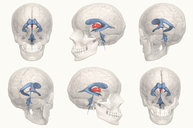
 Third ventricle shown in red | |
 Blue – lateral ventricles Cyan – interventricular foramina (Monro) Yellow – third ventricle Red – cerebral aqueduct (Sylvius) Purple – fourth ventricle Green – continuous with the central canal (apertures to subarachnoid space are not visible) | |
| Details | |
| Identifiers | |
| Latin | ventriculus tertius cerebri |
| MeSH | D020542 |
| NeuroNames | 446 |
| NeuroLex ID | birnlex_714 |
| TA98 | A14.1.08.410 |
| TA2 | 5769 |
| FMA | 78454 |
| Anatomical terms of neuroanatomy | |
The third ventricle is one of the four connected cerebral ventricles of the ventricular system within the mammalian brain. It is a slit-like cavity formed in the diencephalon between the two thalami, in the midline between the right and left lateral ventricles, and is filled with cerebrospinal fluid (CSF).[1]
Running through the third ventricle is the interthalamic adhesion, which contains thalamic neurons and fibers that may connect the two thalami.
- ^ Singh, Vishram (2014). Textbook of Anatomy Head, Neck, and Brain; Volume III (2nd ed.). Elsevier. pp. 386–387. ISBN 9788131237274.