This article needs additional citations for verification. (February 2014) |
| Video-assisted thoracoscopic surgery | |
|---|---|
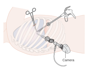
 Diagram showing video assisted thoracoscopy (VATS) | |
| MeSH | D020775 |
Video-assisted thoracoscopic surgery (VATS) is a type of minimally invasive thoracic surgery performed using a small video camera mounted to a fiberoptic thoracoscope (either 5 mm or 10 mm caliber), with or without angulated visualization, which allows the surgeon to see inside the chest by viewing the video images relayed onto a television screen, and perform procedures using elongated surgical instruments. The camera and instruments are inserted into the patient's chest cavity through small incisions in the chest wall, usually via specially designed guiding tubes known as "ports".
VATS procedures are done using either conventional surgical instruments or laparoscopic instruments. Unlike with laparoscopy, carbon dioxide insufflation is not generally required in VATS due to the inherent rigidity of the thoracic cage. However, lung deflation on the side of the operated chest is a must to be able to visualize and pass instruments into the thorax; this is usually effected with a double-lumen endotracheal tube that allows for single-lung ventilation, or a one-side bronchial occlusion delivered via a standard single-lumen tracheal tube.[1]