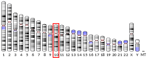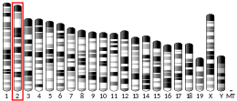
Vimentin is a structural protein that in humans is encoded by the VIM gene. Its name comes from the Latin vimentum which refers to an array of flexible rods.[5]

Vimentin is a type III intermediate filament (IF) protein that is expressed in mesenchymal cells. IF proteins are found in all animal cells[6] as well as bacteria.[7] Intermediate filaments, along with tubulin-based microtubules and actin-based microfilaments, comprises the cytoskeleton. All IF proteins are expressed in a highly developmentally-regulated fashion; vimentin is the major cytoskeletal component of mesenchymal cells. Because of this, vimentin is often used as a marker of mesenchymally-derived cells or cells undergoing an epithelial-to-mesenchymal transition (EMT) during both normal development and metastatic progression.
- ^ a b c GRCh38: Ensembl release 89: ENSG00000026025 – Ensembl, May 2017
- ^ a b c GRCm38: Ensembl release 89: ENSMUSG00000026728 – Ensembl, May 2017
- ^ "Human PubMed Reference:". National Center for Biotechnology Information, U.S. National Library of Medicine.
- ^ "Mouse PubMed Reference:". National Center for Biotechnology Information, U.S. National Library of Medicine.
- ^ Franke WW, Schmid E, Osborn M, Weber K (October 1978). "Different intermediate-sized filaments distinguished by immunofluorescence microscopy". Proceedings of the National Academy of Sciences of the United States of America. 75 (10): 5034–5038. Bibcode:1978PNAS...75.5034F. doi:10.1073/pnas.75.10.5034. PMC 336257. PMID 368806.
- ^ Eriksson JE, Dechat T, Grin B, Helfand B, Mendez M, Pallari HM, et al. (July 2009). "Introducing intermediate filaments: from discovery to disease". The Journal of Clinical Investigation. 119 (7): 1763–1771. doi:10.1172/JCI38339. PMC 2701876. PMID 19587451.
- ^ Cabeen MT, Jacobs-Wagner C (2010). "The bacterial cytoskeleton". Annual Review of Genetics. 44: 365–392. doi:10.1146/annurev-genet-102108-134845. PMID 21047262.


