| Adenomyosis | |
|---|---|
 | |
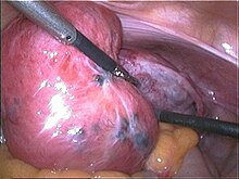
| Adenomyosis uteri seen during laparoscopy: soft and enlarged uterus; the blue spots represent subserous endometriosis. | |
| Specialty | Gynecology |
| Frequency | 20 to 35%.[1] |
Adenomyosis is a medical condition characterized by the growth of cells that proliferate on the inside of the uterus (endometrium) atypically located among the cells of the uterine wall (myometrium),[2] as a result, thickening of the uterus occurs. As well as being misplaced in patients with this condition, endometrial tissue is completely functional. The tissue thickens, sheds and bleeds during every menstrual cycle.[2]
The condition is typically found in women between the ages of 35 and 50, but also affects younger women.[3] Patients with adenomyosis often present with painful menses (dysmenorrhea), profuse menses (menorrhagia), or both. Other possible symptoms are pain during sexual intercourse, chronic pelvic pain and irritation of the urinary bladder.
In adenomyosis, basal endometrium penetrates into hyperplastic myometrial fibers. Unlike the functional layer, the basal layer does not undergo typical cyclic changes with the menstrual cycle.[4][5] Adenomyosis may involve the uterus focally, creating an adenomyoma. With diffuse involvement, the uterus becomes bulky and heavier.[6]
Adenomyosis can be found together with endometriosis; it differs in that patients with endometriosis present endometrial-like tissue located entirely outside the uterus. In endometriosis, the tissue is similar to, but not the same as, the endometrium. The two conditions are found together in many cases yet often occur separately.[7][4] Before being recognized as a distinct condition, adenomyosis was called endometriosis interna. The less-commonly-used term adenomyometritis is a more specific name for the condition, specifying involvement of the uterus.[8][9]
- ^ Cite error: The named reference
statswas invoked but never defined (see the help page). - ^ a b R G, C W (2020). "Adenomyosis". StatPearls [Internet]. PMID 30969690.
- ^ Brosens I, Gordts S, Habiba M, Benagiano G (December 2015). "Uterine Cystic Adenomyosis: A Disease of Younger Women". J Pediatr Adolesc Gynecol. 28 (6): 420–6. doi:10.1016/j.jpag.2014.05.008. PMID 26049940.
- ^ a b Katz VL (2007). Comprehensive gynecology (5th ed.). Philadelphia PA: Mosby Elsevier.
- ^ Leyendecker, G., Herbertz, M., Kunz, G., Mall, G. (2002). "Endometriosis results from the dislocation of basal endometrium". Hum. Reprod. 17 (10): 2725–2736. doi:10.1093/humrep/17.10.2725. PMID 12351554.
- ^ Cite error: The named reference
:0was invoked but never defined (see the help page). - ^ Lazzeri L, Di Giovanni A, Exacoustos C, Tosti C, Pinzauti S, Malzoni M, Petraglia F, Zupi E (August 2014). "Preoperative and Postoperative Clinical and Transvaginal Ultrasound Findings of Adenomyosis in Patients With Deep Infiltrating Endometriosis". Reprod Sci. 21 (8): 1027–1033. doi:10.1177/1933719114522520. PMID 24532217. S2CID 24041889.
- ^ "adenomyometritis" at Dorland's Medical Dictionary
- ^ Matalliotakis I, Kourtis A, Panidis D (2003). "Adenomyosis". Obstetrics and Gynecology Clinics of North America. 30 (1): 63–82, viii. doi:10.1016/S0889-8545(02)00053-0. PMID 12699258.