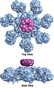
The apoptosome is a large quaternary protein structure formed in the process of apoptosis. Its formation is triggered by the release of cytochrome c from the mitochondria in response to an internal (intrinsic) or external (extrinsic) cell death stimulus. Stimuli can vary from DNA damage and viral infection to developmental cues such as those leading to the degradation of a tadpole's tail.
In mammalian cells, once cytochrome c is released, it binds to the cytosolic protein Apaf-1 to facilitate the formation of an apoptosome. An early biochemical study suggests a two-to-one ratio of cytochrome c to apaf-1 for apoptosome formation. However, recent structural studies suggest the cytochrome c to apaf-1 ratio is one-to-one. It has also been shown that the nucleotide dATP as third component binds to apaf-1, however its exact role is still debated. The mammalian apoptosome had never been crystallized, but a human APAF-1/cytochrome-c apoptosome has been imaged at lower (2 nm) resolution by cryogenic transmission electron microscopy in 2002,[2] revealing a heptameric wheel-like particle with 7-fold symmetry. Recently, a medium resolution (9.5 Ångström) structure of human apoptosome was also solved by cryo-electron microscopy, which allows unambiguous inference for positions of all the APAF-1 domains (CARD, NBARC and WD40) and cytochrome c. There is also now a crystal structure of the monomeric, inactive Apaf-1 subunit (PDB 3SFZ).[1][3]
Once formed, the apoptosome can then recruit and activate the inactive pro-caspase-9. Once activated, this initiator caspase can then activate effector caspases and trigger a cascade of events leading to apoptosis.
- ^ a b Yuan S, Yu X, Topf M, Ludtke SJ, Wang X, Akey CW. "Structure of an apoptosome-procaspase-9 CARD complex." Structure. 2010 May;18(5):571-83.
- ^ Cite error: The named reference
Threewas invoked but never defined (see the help page). - ^ T. F. Reubold; S. Wohlgemuth; S. Eschenburg (2011). "Crystal structure of full-length Apaf-1: how the death signal is relayed in the mitochondrial pathway of apoptosis". Structure. 19 (8): 1074–1083. doi:10.1016/j.str.2011.05.013. PMID 21827944.