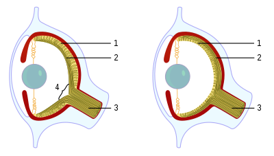
Cephalopods, as active marine predators, possess sensory organs specialized for use in aquatic conditions.[1] They have a camera-type eye which consists of an iris, a circular lens, vitreous cavity (eye gel), pigment cells, and photoreceptor cells that translate light from the light-sensitive retina into nerve signals which travel along the optic nerve to the brain.[2] For the past 140 years, the camera-type cephalopod eye has been compared with the vertebrate eye as an example of convergent evolution, where both types of organisms have independently evolved the camera-eye trait and both share similar functionality. Contention exists on whether this is truly convergent evolution or parallel evolution.[3] Unlike the vertebrate camera eye, the cephalopods' form as invaginations of the body surface (rather than outgrowths of the brain), and consequently the cornea lies over the top of the eye as opposed to being a structural part of the eye.[4] Unlike the vertebrate eye, a cephalopod eye is focused through movement, much like the lens of a camera or telescope, rather than changing shape as the lens in the human eye does. The eye is approximately spherical, as is the lens, which is fully internal.[5]
Cephalopods' eyes develop in such a way that they have retinal axons that pass over the back of the retina, so the optic nerve does not have to pass through the photoreceptor layer to exit the eye and do not have the natural, central, physiological blind spot of vertebrates.[2]
The crystallins used in the lens appear to have developed independently from vertebrate crystallins, suggesting a homoplasious origin of the lens.[6]
Most cephalopods possess complex extraocular muscle systems that allow for very fine control over the gross positioning of the eyes. Octopuses possess an autonomic response that maintains the orientation of their pupils such that they are always horizontal.[1]
- ^ a b Budelmann BU. "Cephalopod sense organs, nerves and the brain: Adaptations for high performance and life style." Marine and Freshwater Behavior and Physiology. Vol 25, Issue 1-3, Page 13-33.
- ^ a b Serb, Jeanne M. (2008). "Toward Developing Models to Study the Disease, Ecology, and Evolution of the Eye in Mollusca*" (PDF). American Malacological Bulletin. 26 (1–2): 3–18. doi:10.4003/006.026.0202. S2CID 1557944. Archived from the original (PDF) on 2014-12-18. Retrieved 2014-11-18.
- ^ Serb, J.; Eernisse, D. (2008). "Charting Evolution's Trajectory: Using Molluscan Eye Diversity to Understand Parallel and Convergent Evolution". Evolution: Education & Outreach. 1 (4): 439–447. doi:10.1007/s12052-008-0084-1.
- ^ Hanke, Frederike D.; Kelber, Almut (2020-01-14). "The Eye of the Common Octopus (Octopus vulgaris)". Frontiers in Physiology. 10: 1637. doi:10.3389/fphys.2019.01637. ISSN 1664-042X. PMC 6971404. PMID 32009987.
- ^ Yamamoto, M. (Feb 1985). "Ontogeny of the visual system in the cuttlefish, Sepiella japonica. I. Morphological differentiation of the visual cell". The Journal of Comparative Neurology. 232 (3): 347–361. doi:10.1002/cne.902320307. ISSN 0021-9967. PMID 2857734. S2CID 24458056.
- ^ SAMIR K. BRAHMA1 (1978). "Ontogeny of lens crystallins in marine cephalopods" (PDF). Embryol. Exp. Morph. 46 (1): 111–118. PMID 359745.
{{cite journal}}: CS1 maint: numeric names: authors list (link)
