Free fatty acid receptor 3 (FFAR3, also termed GPR41) protein is a G protein coupled receptor (i.e., GPR or GPCR) that in humans is encoded by the FFAR3 gene (i.e., GPR41 gene).[5] GPRs reside on cell surfaces, bind specific signaling molecules, and thereby are activated to trigger certain functional responses in their parent cells. FFAR3 is a member of the free fatty acid receptor group of GPRs that includes FFAR1 (i.e., GPR40), FFAR2 (i.e., GPR43), and FFAR4 (i.e., GPR120).[6] All of these FFARs are activated by fatty acids. FFAR3 and FFAR2 are activated by certain short-chain fatty acids (SC-FAs), i.e., fatty acids consisting of 2 to 6 carbon atoms[7] whereas FFFAR1 and FFAR4 are activated by certain fatty acids that are 6 to more than 21 carbon atoms long.[8][9][10] Hydroxycarboxylic acid receptor 2 is also activated by a SC-FA that activate FFAR3, i.e., butyric acid.[11]
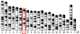
The human FFAR3 gene is located next to the FFAR2 gene at locus 13.12 on the long (i.e., "q") arm of chromosome 19 (location abbreviated as 19q13.12). The human FFAR3 and FFAR2 proteins consist of 346 and 330 amino acids, respectively,[12] and share about a 40% amino acid sequence homology.[13] The two FFARs have been found to form a heteromer complex (i.e., FFAR3 and FFAR2 bind to each other and are activated together by a SC-FA) in human monocytes, macrophages, and the immortalized embryonic kidney cells, HEK 293 cells. When stimulated by a SC-FA, the cells expressing both FFAR3 and FFAR2 may form this heterodimer and thereby activate cell signaling pathways and mount responses that differ from those of cells expressing only one of these FFARs.[14] The formation of GPR43-GPR41 heterodimers has not been evaluated in most studies and may explain otherwise conflicting results on the roles of FFAR3 and FFAR2 in cell function.[10][15][16] Furthermore, SC-FAs can alter the function of cells independently of FFAR3 and FFAR2 by altering the activity of cellular histone deacetylases which regulate the transcription of various genes or by altering metabolic pathways which alter cell functions.[17][18] Given these alternate ways for SC-FAs to activate cells as wells at the ability of SC-FAs to activate FFAR2 or, in the case of butyric acid, hydroxycarboxylic acid receptor 2, the studies reported here focus on those showing that the examined action(s) of an SC-FA is absent or reduced in cells, tissues, or animals that have no or reduced FFAR3 activity due respectively to knockout (i.e., removal or inactivation) or knockdown (i.e., reduction) of the FFAR3 protein gene, i.e., the Ffar3 gene in animals or FFAR3 gene in humans.
Certain bacteria in the gastrointestinal tract ferment fecal fiber into SC-FAs and excrete them as waste products. The excreted SC-FAs enter the gastrointestinal walls, diffuse into the portal venous system, and ultimately flow into the systemic circulation. During this passage, they can activate the FFAR3 on cells in the intestinal wall as well as throughout the body.[19] This activation may: suppress the appetite for food and thereby reduce overeating and the development of obesity;[20][21] inhibit the liver's accumulation of fatty acids and thereby the development of fatty liver diseases;[22] decrease blood pressure and thereby the development of hypertension and hypertension-related cardiac diseases;[23] modulate insulin secretion and thereby the development and/or symptoms of type 2 diabetes;[24] reduce heart rate and blood plasma norepinephrine levels and thereby lower total body energy expenditures;[19] and suppress or delay the development of allergic asthma.[25]
The specific types of bacteria in the intestines can be modified to increase the number which make SC-FAs by using foods that stimulate the growth of these bacteria (i.e., prebiotics), preparations of SC-FA-producing bacteria (i.e., probiotics), or both methods (see synbiotics).[26] Individuals with disorders that are associated with low levels of the SC-FA-producing intestinal bacteria may show improvements in their conditions when treated with prebiotics, probiotics, or synbiotics while individuals with disorders associated with high levels of SC-FAs may show improvements in their conditions when treated with methods, e.g., antibiotics, that reduce the intestinal levels of these bacteria.[19][27] (For information on these treatments see Disorders treated by probiotics and Disorders treated by prebiotics). In addition, drugs are being tested for their ability to act more usefully, potently, and effectively than SC-FAs in stimulating or inhibiting FFAR3 and thereby for treating the disoders that are inhibited or stimulated, respectively, by SC-FAs.[28]
- ^ a b c GRCh38: Ensembl release 89: ENSG00000185897 – Ensembl, May 2017
- ^ a b c GRCm38: Ensembl release 89: ENSMUSG00000019429 – Ensembl, May 2017
- ^ "Human PubMed Reference:". National Center for Biotechnology Information, U.S. National Library of Medicine.
- ^ "Mouse PubMed Reference:". National Center for Biotechnology Information, U.S. National Library of Medicine.
- ^ "Entrez Gene: FFAR3 free fatty acid receptor 3".
- ^ Sawzdargo M, George SR, Nguyen T, Xu S, Kolakowski LF, O'Dowd BF (October 1997). "A cluster of four novel human G protein-coupled receptor genes occurring in close proximity to CD22 gene on chromosome 19q13.1". Biochemical and Biophysical Research Communications. 239 (2): 543–7. doi:10.1006/bbrc.1997.7513. PMID 9344866.
- ^ Karmokar PF, Moniri NH (December 2022). "Oncogenic signaling of the free-fatty acid receptors FFA1 and FFA4 in human breast carcinoma cells". Biochemical Pharmacology. 206: 115328. doi:10.1016/j.bcp.2022.115328. PMID 36309079. S2CID 253174629.
- ^ Briscoe CP, Tadayyon M, Andrews JL, Benson WG, Chambers JK, Eilert MM, Ellis C, Elshourbagy NA, Goetz AS, Minnick DT, Murdock PR, Sauls HR, Shabon U, Spinage LD, Strum JC, Szekeres PG, Tan KB, Way JM, Ignar DM, Wilson S, Muir AI (March 2003). "The orphan G protein-coupled receptor GPR40 is activated by medium and long chain fatty acids". The Journal of Biological Chemistry. 278 (13): 11303–11. doi:10.1074/jbc.M211495200. PMID 12496284.
- ^ Tunaru S, Bonnavion R, Brandenburger I, Preussner J, Thomas D, Scholich K, Offermanns S (January 2018). "20-HETE promotes glucose-stimulated insulin secretion in an autocrine manner through FFAR1". Nature Communications. 9 (1): 177. Bibcode:2018NatCo...9..177T. doi:10.1038/s41467-017-02539-4. PMC 5766607. PMID 29330456.
- ^ a b Grundmann M, Bender E, Schamberger J, Eitner F (February 2021). "Pharmacology of Free Falsoatty Acid Receptors and Their Allosteric Modulators". International Journal of Molecular Sciences. 22 (4). doi:10.3390/ijms22041763. PMC 7916689. PMID 33578942.
- ^ Offermanns S (March 2017). "Hydroxy-Carboxylic Acid Receptor Actions in Metabolism". Trends in Endocrinology and Metabolism. 28 (3): 227–236. doi:10.1016/j.tem.2016.11.007. PMID 28087125. S2CID 39660018.
- ^ Mishra SP, Karunakar P, Taraphder S, Yadav H (June 2020). "Free Fatty Acid Receptors 2 and 3 as Microbial Metabolite Sensors to Shape Host Health: Pharmacophysiological View". Biomedicines. 8 (6): 154. doi:10.3390/biomedicines8060154. PMC 7344995. PMID 32521775.
- ^ Secor JD, Fligor SC, Tsikis ST, Yu LJ, Puder M (2021). "Free Fatty Acid Receptors as Mediators and Therapeutic Targets in Liver Disease". Frontiers in Physiology. 12: 656441. doi:10.3389/fphys.2021.656441. PMC 8058363. PMID 33897464.
- ^ Ang Z, Xiong D, Wu M, Ding JL (January 2018). "FFAR2-FFAR3 receptor heteromerization modulates short-chain fatty acid sensing". FASEB Journal. 32 (1): 289–303. doi:10.1096/fj.201700252RR. PMC 5731126. PMID 28883043.
- ^ Martin-Gallausiaux C, Marinelli L, Blottière HM, Larraufie P, Lapaque N (February 2021). "SCFA: mechanisms and functional importance in the gut". The Proceedings of the Nutrition Society. 80 (1): 37–49. doi:10.1017/S0029665120006916. PMID 32238208. S2CID 214772999.
- ^ Ang Z, Er JZ, Tan NS, Lu J, Liou YC, Grosse J, Ding JL (September 2016). "Human and mouse monocytes display distinct signalling and cytokine profiles upon stimulation with FFAR2/FFAR3 short-chain fatty acid receptor agonists". Scientific Reports. 6: 34145. Bibcode:2016NatSR...634145A. doi:10.1038/srep34145. PMC 5036191. PMID 27667443.
- ^ Carretta MD, Quiroga J, López R, Hidalgo MA, Burgos RA (2021). "Participation of Short-Chain Fatty Acids and Their Receptors in Gut Inflammation and Colon Cancer". Frontiers in Physiology. 12: 662739. doi:10.3389/fphys.2021.662739. PMC 8060628. PMID 33897470.
- ^ Tan JK, Macia L, Mackay CR (February 2023). "Dietary fiber and SCFAs in the regulation of mucosal immunity". The Journal of Allergy and Clinical Immunology. 151 (2): 361–370. doi:10.1016/j.jaci.2022.11.007. PMID 36543697. S2CID 254918066.
- ^ a b c Ikeda T, Nishida A, Yamano M, Kimura I (November 2022). "Short-chain fatty acid receptors and gut microbiota as therapeutic targets in metabolic, immune, and neurological diseases". Pharmacology & Therapeutics. 239: 108273. doi:10.1016/j.pharmthera.2022.108273. PMID 36057320. S2CID 251992642.
- ^ Obradovic M, Sudar-Milovanovic E, Soskic S, Essack M, Arya S, Stewart AJ, Gojobori T, Isenovic ER (2021). "Leptin and Obesity: Role and Clinical Implication". Frontiers in Endocrinology. 12: 585887. doi:10.3389/fendo.2021.585887. PMC 8167040. PMID 34084149.
- ^ Navalón-Monllor V, Soriano-Romaní L, Silva M, de Las Hazas ML, Hernando-Quintana N, Suárez Diéguez T, Esteve PM, Nieto JA (August 2023). "Microbiota dysbiosis caused by dietetic patterns as a promoter of Alzheimer's disease through metabolic syndrome mechanisms". Food & Function. 14 (16): 7317–7334. doi:10.1039/d3fo01257c. PMID 37470232. S2CID 259996464.
- ^ Shimizu H, Masujima Y, Ushiroda C, Mizushima R, Taira S, Ohue-Kitano R, Kimura I (November 2019). "Dietary short-chain fatty acid intake improves the hepatic metabolic condition via FFAR3". Scientific Reports. 9 (1): 16574. Bibcode:2019NatSR...916574S. doi:10.1038/s41598-019-53242-x. PMC 6851370. PMID 31719611.
- ^ Pluznick JL, Protzko RJ, Gevorgyan H, Peterlin Z, Sipos A, Han J, Brunet I, Wan LX, Rey F, Wang T, Firestein SJ, Yanagisawa M, Gordon JI, Eichmann A, Peti-Peterdi J, Caplan MJ (March 2013). "Olfactory receptor responding to gut microbiota-derived signals plays a role in renin secretion and blood pressure regulation". Proceedings of the National Academy of Sciences of the United States of America. 110 (11): 4410–5. Bibcode:2013PNAS..110.4410P. doi:10.1073/pnas.1215927110. PMC 3600440. PMID 23401498.
- ^ Ghislain J, Poitout V (March 2021). "Targeting lipid GPCRs to treat type2 diabetes mellitus - progress and challenges". Nature Reviews. Endocrinology. 17 (3): 162–175. doi:10.1038/s41574-020-00459-w. PMID 33495605. S2CID 231695737.
- ^ Yuan G, Wen S, Zhong X, Yang X, Xie L, Wu X, Li X (2023). "Inulin alleviates offspring asthma by altering maternal intestinal microbiome composition to increase short-chain fatty acids". PLOS ONE. 18 (4): e0283105. Bibcode:2023PLoSO..1883105Y. doi:10.1371/journal.pone.0283105. PMC 10072493. PMID 37014871.
- ^ Kim YA, Keogh JB, Clifton PM (June 2018). "Probiotics, prebiotics, synbiotics and insulin sensitivity". Nutrition Research Reviews. 31 (1): 35–51. doi:10.1017/S095442241700018X. PMID 29037268.
- ^ Cite error: The named reference
pmid31487233was invoked but never defined (see the help page). - ^ Loona DP, Das B, Kaur R, Kumar R, Yadav AK (2023). "Free Fatty Acid Receptors (FFARs): Emerging Therapeutic Targets for the Management of Diabetes Mellitus". Current Medicinal Chemistry. 30 (30): 3404–3440. doi:10.2174/0929867329666220927113614. PMID 36173072. S2CID 252598831.


