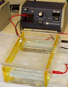
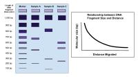
 Gel electrophoresis apparatus – an agarose gel is placed in this buffer-filled box and an electric current is applied via the power supply to the rear. The negative terminal is at the far end (black wire), so DNA migrates toward the positively charged anode(red wire). This occurs because phosphate groups found in the DNA fragments possess a negative charge which is repelled by the negatively charged cathode and are attracted to the positively charged anode. | |
| Classification | Electrophoresis |
|---|---|
| Other techniques | |
| Related | Capillary electrophoresis SDS-PAGE Two-dimensional gel electrophoresis Temperature gradient gel electrophoresis |


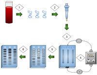
- DNA is extracted.
- Isolation and amplification of DNA.
- DNA added to the gel wells.
- Electric current applied to the gel.
- DNA bands are separated by size.
- DNA bands are stained.
Gel electrophoresis is a method for separation and analysis of biomacromolecules (DNA, RNA, proteins, etc.) and their fragments, based on their size and charge. It is used in clinical chemistry to separate proteins by charge or size (IEF agarose, essentially size independent) and in biochemistry and molecular biology to separate a mixed population of DNA and RNA fragments by length, to estimate the size of DNA and RNA fragments or to separate proteins by charge.[1]
Nucleic acid molecules are separated by applying an electric field to move the negatively charged molecules through a matrix of agarose or other substances. Shorter molecules move faster and migrate farther than longer ones because shorter molecules migrate more easily through the pores of the gel. This phenomenon is called sieving.[2] Proteins are separated by the charge in agarose because the pores of the gel are too large to sieve proteins. Gel electrophoresis can also be used for the separation of nanoparticles.
Gel electrophoresis uses a gel as an anticonvective medium or sieving medium during electrophoresis, the movement of a charged particle in an electric current. Gels suppress the thermal convection caused by the application of the electric field, and can also act as a sieving medium, slowing the passage of molecules; gels can also simply serve to maintain the finished separation so that a post electrophoresis stain can be applied.[3] DNA gel electrophoresis is usually performed for analytical purposes, often after amplification of DNA via polymerase chain reaction (PCR), but may be used as a preparative technique prior to use of other methods such as mass spectrometry, RFLP, PCR, cloning, DNA sequencing, or Southern blotting for further characterization.
- ^ Kryndushkin DS; Alexandrov IM; Ter-Avanesyan MD; Kushnirov VV (2003). "Yeast [PSI+] prion aggregates are formed by small Sup35 polymers fragmented by Hsp104". J Biol Chem. 278 (49): 49636–43. doi:10.1074/jbc.M307996200. PMID 14507919.
- ^ Sambrook, Joseph (2001). Molecular cloning : a laboratory manual (in Spanish). Cold Spring Harbor, N.Y: Cold Spring Harbor Laboratory Press. ISBN 978-0-87969-576-7. OCLC 45015638.
- ^ Berg, Jeremy (2002). Biochemistry (in Estonian). New York: W.H. Freeman. ISBN 978-0-7167-4955-4. OCLC 48055706.