| Hemidesmosome | |
|---|---|
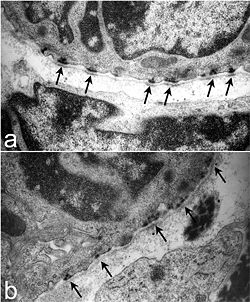
 Ultrastructure of tracheal hemidesmosomes in mice. In a normal mouse (a) there are well-defined, organized hemidesmosomes with darkened areas in the lamina densa abutting the hemidesmosome (arrows). In contrast, hemidesmosomes in Lamc2 -/- tracheas (b) are less organized, the intracellular component is more diffuse, and the lamina densa directly below the hemidesmosomal areas lacks the electron density seen in the littermate control (arrows). From Nguyen et al., 2006.[1] | |
| Details | |
| Identifiers | |
| Latin | hemidesmosoma |
| MeSH | D022002 |
| TH | H1.00.01.1.02029 |
| FMA | 67415 |
| Anatomical terminology | |
Hemidesmosomes are very small stud-like structures found in keratinocytes of the epidermis of skin that attach to the extracellular matrix. They are similar in form to desmosomes when visualized by electron microscopy; however, desmosomes attach to adjacent cells. Hemidesmosomes are also comparable to focal adhesions, as they both attach cells to the extracellular matrix. Instead of desmogleins and desmocollins in the extracellular space, hemidesmosomes utilize integrins. Hemidesmosomes are found in epithelial cells connecting the basal epithelial cells to the lamina lucida, which is part of the basal lamina.[2] Hemidesmosomes are also involved in signaling pathways, such as keratinocyte migration or carcinoma cell intrusion.[3]
- ^ Nguyen NM, Pulkkinen L, Schlueter JA, Meneguzzi G, Uitto J, Senior RM (2006). "Lung development in laminin γ2 deficiency: abnormal tracheal hemidesmosomes with normal branching morphogenesis and epithelial differentiation". Respir. Res. 7 (1): 28. doi:10.1186/1465-9921-7-28. PMC 1386662. PMID 16483354.
- ^ Walko, Gernot; Castañón, Maria J.; Wiche, Gerhard (May 2015). "Molecular architecture and function of the hemidesmosome". Cell and Tissue Research. 360 (2): 363–378. doi:10.1007/s00441-014-2061-z. ISSN 1432-0878. PMC 4544487. PMID 25487405.
- ^ Wilhelmsen, Kevin; Litjens, Sandy H. M.; Sonnenberg, Arnoud (April 2006). "Multiple functions of the integrin alpha6beta4 in epidermal homeostasis and tumorigenesis". Molecular and Cellular Biology. 26 (8): 2877–2886. doi:10.1128/MCB.26.8.2877-2886.2006. ISSN 0270-7306. PMC 1446957. PMID 16581764.