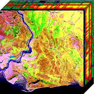
Hyperspectral imaging collects and processes information from across the electromagnetic spectrum.[1] The goal of hyperspectral imaging is to obtain the spectrum for each pixel in the image of a scene, with the purpose of finding objects, identifying materials, or detecting processes.[2][3] There are three general types of spectral imagers. There are push broom scanners and the related whisk broom scanners (spatial scanning), which read images over time, band sequential scanners (spectral scanning), which acquire images of an area at different wavelengths, and snapshot hyperspectral imagers, which uses a staring array to generate an image in an instant.
Whereas the human eye sees color of visible light in mostly three bands (long wavelengths, perceived as red; medium wavelengths, perceived as green; and short wavelengths, perceived as blue), spectral imaging divides the spectrum into many more bands. This technique of dividing images into bands can be extended beyond the visible. In hyperspectral imaging, the recorded spectra have fine wavelength resolution and cover a wide range of wavelengths. Hyperspectral imaging measures continuous spectral bands, as opposed to multiband imaging which measures spaced spectral bands.[4]
Engineers build hyperspectral sensors and processing systems for applications in astronomy, agriculture, molecular biology, biomedical imaging, geosciences, physics, and surveillance. Hyperspectral sensors look at objects using a vast portion of the electromagnetic spectrum. Certain objects leave unique "fingerprints" in the electromagnetic spectrum. Known as spectral signatures, these "fingerprints" enable identification of the materials that make up a scanned object. For example, a spectral signature for oil helps geologists find new oil fields.[5]
- ^ Chilton, Alexander (2013-10-07). "The Working Principle and Key Applications of Infrared Sensors". AZoSensors. Retrieved 2020-07-11.
- ^ Chein-I Chang (31 July 2003). Hyperspectral Imaging: Techniques for Spectral Detection and Classification. Springer Science & Business Media. ISBN 978-0-306-47483-5.
- ^ Hans Grahn; Paul Geladi (27 September 2007). Techniques and Applications of Hyperspectral Image Analysis. John Wiley & Sons. ISBN 978-0-470-01087-7.
- ^ Hagen, Nathan; Kudenov, Michael W. (2013). "Review of snapshot spectral imaging technologies" (PDF). Optical Engineering. 52 (9): 090901. Bibcode:2013OptEn..52i0901H. doi:10.1117/1.OE.52.9.090901. S2CID 215807781.
- ^ Lu, G; Fei, B (January 2014). "Medical Hyperspectral Imaging: a review". Journal of Biomedical Optics. 19 (1): 10901. Bibcode:2014JBO....19a0901L. doi:10.1117/1.JBO.19.1.010901. PMC 3895860. PMID 24441941.