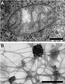
Immunogold labeling or immunogold staining (IGS) is a staining technique used in electron microscopy.[2] This staining technique is an equivalent of the indirect immunofluorescence technique for visible light. Colloidal gold particles are most often attached to secondary antibodies which are in turn attached to primary antibodies designed to bind a specific antigen or other cell component. Gold is used for its high electron density which increases electron scatter to give high contrast 'dark spots'.[3]
First used in 1971, immunogold labeling has been applied to both transmission electron microscopy and scanning electron microscopy, as well as brightfield microscopy. The labeling technique can be adapted to distinguish multiple objects by using differently-sized gold particles.
Immunogold labeling can introduce artifacts, as the gold particles reside some distance from the labelled object and very thin sectioning is required during sample preparation.[3]
- ^ Iborra FJ, Kimura H, Cook PR (2004). "The functional organization of mitochondrial genomes in human cells". BMC Biol. 2: 9. doi:10.1186/1741-7007-2-9. PMC 425603. PMID 15157274.
- ^ "Immunogold Labeling in Scanning Electron Microscopy". Archived from the original on 2014-02-06. Retrieved 2010-07-08.
- ^ a b Alberts, Bruce; et al. (2008). Molecular biology of the cell (5th ed.). New York: Garland Science. ISBN 978-0-8153-4106-2.