| Intraparietal sulcus | |
|---|---|
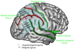
 Lateral surface of left cerebral hemisphere, viewed from the side. (Intraparietal sulcus visible at upper right, running horizontally.) | |
 Right cerebral hemisphere, viewed from the side. The region colored in blue is parietal lobe of the human brain. Intraparietal sulcus runs horizontally at the middle of the parietal lobe. | |
| Details | |
| Part of | Parietal lobe |
| Identifiers | |
| Latin | sulcus intraparietalis |
| Acronym(s) | IPS |
| NeuroNames | 97 |
| NeuroLex ID | birnlex_4031 |
| TA98 | A14.1.09.127 |
| TA2 | 5475 |
| FMA | 83772 |
| Anatomical terms of neuroanatomy | |
The intraparietal sulcus (IPS) is located on the lateral surface of the parietal lobe, and consists of an oblique and a horizontal portion. The IPS contains a series of functionally distinct subregions that have been intensively investigated using both single cell neurophysiology in primates[1][2] and human functional neuroimaging.[3] Its principal functions are related to perceptual-motor coordination (e.g., directing eye movements and reaching) and visual attention, which allows for visually-guided pointing, grasping, and object manipulation that can produce a desired effect.
The intraparietal sulcus (IPS) plays a pivotal role in multisensory integration, particularly in linking visual and tactile information to guide complex motor actions. Beyond its established roles in numerical cognition and spatial attention, the IPS has emerged as a critical player in tool use and manipulation.[4]
The IPS is also thought to play a role in other functions, including processing symbolic numerical information,[5] visuospatial working memory,[6] decision-making,[7] and interpreting the intent of others.[8][unreliable medical source?]
- ^ Colby C.E.; Goldberg M.E. (1999). "Space and attention in parietal cortex". Annual Review of Neuroscience. 22: 319–349. doi:10.1146/annurev.neuro.22.1.319. PMID 10202542.
- ^ Andersen R.A. (1989). "Visual and eye movement functions of the posterior parietal cortex" (PDF). Annual Review of Neuroscience. 12: 377–403. doi:10.1146/annurev.ne.12.030189.002113. PMID 2648954.
- ^ Culham, J.C.; Nancy G. Kanwisher (April 2001). "Neuroimaging of cognitive functions in human parietal cortex". Current Opinion in Neurobiology. 11 (2): 157–163. doi:10.1016/S0959-4388(00)00191-4. PMID 11301234. S2CID 13907037.
- ^ Swisher, Jascha D.; Halko, Mark A.; Merabet, Lotfi B.; McMains, Stephanie A.; Somers, David C. (2007-05-16). "Visual Topography of Human Intraparietal Sulcus". Journal of Neuroscience. 27 (20): 5326–5337. doi:10.1523/JNEUROSCI.0991-07.2007. ISSN 0270-6474. PMID 17507555.
- ^ Cantlon J, Brannon E, Carter E, Pelphrey K (2006). "Functional imaging of numerical processing in adults and 4-y-old children". PLOS Biol. 4 (5): e125. doi:10.1371/journal.pbio.0040125. PMC 1431577. PMID 16594732.
- ^ Todd JJ, Marois R (2004). "Capacity limit of visual short-term memory in human posterior parietal cortex". Nature. 428 (6984): 751–754. Bibcode:2004Natur.428..751T. doi:10.1038/nature02466. PMID 15085133. S2CID 4415712.
- ^ Valdebenito-Oyarzo, Gabriela; Martínez-Molina, María Paz; Soto-Icaza, Patricia; Zamorano, Francisco; Figueroa-Vargas, Alejandra; Larraín-Valenzuela, Josefina; Stecher, Ximena; Salinas, César; Bastin, Julien; Valero-Cabré, Antoni; Polania, Rafael; Billeke, Pablo (10 January 2024). "The parietal cortex has a causal role in ambiguity computations in humans". PLOS Biology. 22 (1): e3002452. doi:10.1371/journal.pbio.3002452. PMC 10824459. PMID 38198502.
- ^ Grafton, Hamilton (2006). "Dartmouth Study Finds How The Brain Interprets The Intent Of Others". Science Daily.