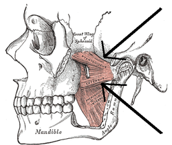
| Lateral pterygoid muscle | |
|---|---|
 The Pterygoidei; the zygomatic arch and a portion of the ramus of the mandible have been removed (labeled as "pterygoideus externus", visible in pink at center) | |
 Sagittal section of the temporomandibular joint (labeled as "pterygoideus externus", visible in gray at bottom left) | |
| Details | |
| Origin | Superior head: infratemporal surface of sphenoid bone. Inferior head: lateral pterygoid plate |
| Insertion | Superior head: anterior side of the mandibular condyle. Inferior head: pterygoid fovea |
| Artery | Pterygoid branches of maxillary artery |
| Nerve | Lateral pterygoid nerve from mandibular nerve |
| Actions | Depresses and protrudes mandible, side to side movement of mandible |
| Identifiers | |
| Latin | musculus pterygoideus lateralis, musculus pterygoideus externus |
| TA98 | A04.1.04.006 |
| TA2 | 2109 |
| FMA | 49015 |
| Anatomical terms of muscle | |
The lateral pterygoid muscle (or external pterygoid muscle) is a muscle of mastication. It has two heads. It lies superior to the medial pterygoid muscle. It is supplied by pterygoid branches of the maxillary artery, and the lateral pterygoid nerve (from the mandibular nerve, CN V3). It depresses and protrudes the mandible. When each muscle works independently, they can move the mandible side to side.