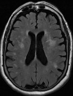


Leukoaraiosis is a particular abnormal change in appearance of white matter near the lateral ventricles. It is often seen in aged individuals, but sometimes in young adults.[1][2] On MRI, leukoaraiosis changes appear as white matter hyperintensities (WMHs) in T2 FLAIR images.[3][4] On CT scans, leukoaraiosis appears as hypodense periventricular white-matter lesions.[5]
The term "leukoaraiosis" was coined in 1986[6][7] by Hachinski, Potter, and Merskey as a descriptive term for rarefaction ("araiosis") of the white matter, showing up as decreased density on CT and increased signal intensity on T2/FLAIR sequences (white matter hyperintensities) performed as part of MRI brain scans.
These white matter changes are also commonly referred to as periventricular white matter disease, or white matter hyperintensities (WMH), due to their bright white appearance on T2 MRI scans. Many patients can have leukoaraiosis without any associated clinical abnormality. However, underlying vascular mechanisms are suspected to be the cause of the imaging findings. Hypertension, smoking, diabetes,[3] hyperhomocysteinemia, and heart diseases are all risk factors for leukoaraiosis.
Leukoaraiosis has been reported to be an initial stage of Binswanger's disease but this evolution does not always happen.
- ^ Putaala J., Kurkinen M., Tarvos V., Salonen O., Kaste M., Tatlisumak T. (2009). "Silent brain infarcts and leukoaraiosis in young adults with first-ever ischemic stroke". Neurology. 72 (21): 1823–1829. doi:10.1212/WNL.0b013e3181a711df. PMID 19470964. S2CID 593328.
{{cite journal}}: CS1 maint: multiple names: authors list (link) - ^ Aik Kah, Tan (2018). "CuRRL Syndrome: A Case Series" (PDF). Acta Scientific Ophthalmology. 1 (3): 9–13.
- ^ a b Habes M, Erus G, Toledo JB, Zhang T, Bryan N, Launer LJ, Rosseel Y, Janowitz D, Doshi J, Van der Auwera S, von Sarnowski B, Hegenscheid K, Hosten N, Homuth G, Völzke H, Schminke U, Hoffmann W, Grabe H, Davatzikos C (2016). "White matter hyperintensities and imaging patterns of brain ageing in the general population". Brain. 139 (Pt 4): 1164–79. doi:10.1093/brain/aww008. PMC 5006227. PMID 26912649.
- ^ Yan, Shenqiang; Wan, Jinping; Zhang, Xuting; Tong, Lusha; Zhao, Song; Sun, Jianzhong; Lin, Yuehan; Shen, Chunhong; Lou, Min (2014). "Increased Visibility of Deep Medullary Veins in Leukoaraiosis: A 3-T MRI Study". Frontiers in Aging Neuroscience. 6: 144. doi:10.3389/fnagi.2014.00144. PMC 4074703. PMID 25071553.
- ^ Kobari M, Meyer JS, Ichijo M, Oravez WT (1990). "Leukoaraiosis: correlation of MR and CT findings with blood flow, atrophy, and cognition". AJNR Am J Neuroradiol. 11 (2): 273–81. PMC 8334682. PMID 2107711.
- ^ Hachinski, VC; Potter, P; Merskey, H (1986). "Leuko-araiosis: An ancient term for a new problem". The Canadian Journal of Neurological Sciences. 13 (4 Suppl): 533–34. doi:10.1017/S0317167100037264. PMID 3791068. S2CID 38151019.
- ^ Hachinski, V. C.; Potter, P.; Merskey, H. (1987). "Leuko-Araiosis". Archives of Neurology. 44 (1): 21–23. doi:10.1001/archneur.1987.00520130013009. PMID 3800716.