| Macula | |
|---|---|
 | |
| Details | |
| Part of | Retina of human eye |
| System | Visual system |
| Identifiers | |
| Latin | macula lutea |
| MeSH | D008266 |
| TA98 | A15.2.04.021 |
| TA2 | 6784 |
| FMA | 58637 |
| Anatomical terminology | |
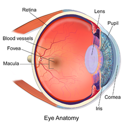
The macula (/ˈmakjʊlə/)[1] or macula lutea is an oval-shaped pigmented area in the center of the retina of the human eye and in other animals. The macula in humans has a diameter of around 5.5 mm (0.22 in) and is subdivided into the umbo, foveola, foveal avascular zone, fovea, parafovea, and perifovea areas.[2]
The anatomical macula at a size of 5.5 mm (0.22 in) is much larger than the clinical macula which, at a size of 1.5 mm (0.059 in), corresponds to the anatomical fovea.[3][4][5]
The macula is responsible for the central, high-resolution, color vision that is possible in good light. This kind of vision is impaired if the macula is damaged, as in macular degeneration. The clinical macula is seen when viewed from the pupil, as in ophthalmoscopy or retinal photography.
The term macula lutea comes from Latin macula, "spot", and lutea, "yellow".
- ^ "MACULA | Meaning & Definition for UK English". Lexico.com. Archived from the original on 3 August 2020. Retrieved 24 August 2022.
- ^ "Interpretation of Stereo Ocular Angiography : Retinal and Choroidal Anatomy". Project Orbis International. Archived from the original on 19 December 2014. Retrieved 11 October 2014.
- ^ Yanoff, Myron (2009). Ocular Pathology. Elsevier Health Sciences. p. 393. ISBN 978-0-323-04232-1. Retrieved 7 November 2014.
- ^ Small, R.G. (1994). The Clinical Handbook of Ophthalmology. CRC Press. p. 134. ISBN 978-1-85070-584-0. Retrieved 7 November 2014.
- ^ Peyman, Gholam A.; Meffert, Stephen A.; Chou, Famin; Mandi D. Conway (2000). Vitreoretinal Surgical Techniques. CRC Press. p. 6. ISBN 978-1-85317-585-5. Retrieved 7 November 2014.