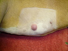
A mastocytoma in dogs (or mast cell tumor in dogs) is a neoplasm (neoplasia) originating from mast cells in the domestic dog, which occurs mainly in the skin and subcutis. Mastocytoma are not only extremely common in dogs, but also tend to be much more malignant in them than in other animal species. The average survival time for malignant tumors is only four months, whereas for benign tumors it is over two years.

Mast cells are cells of the immune system that play a role in the innate immune response. They produce a number of biologically active substances, including primarily histamine. Mastocytoma account for about one-fifth of all skin tumors in dogs. They present as nodules or raised patches, and about one-fifth of affected animals have ulcers and bleeding in the stomach and duodenum. Metastasis in malignant mastocytoma occur primarily in lymph nodes, liver, spleen, and bone marrow. Any lump in the skin or subcutaneous tissue can be a mastocytoma. Detection is only possible by taking tissue with a fine needle (fine needle biopsy) followed by staining and microscopic examination (cytopathology).
Although the classifications according to the clinical appearances and cell appearance in cytodiagnostics give indications of the biological behavior (benign or malignant) and thus the prospect of cure, this tumor disease is unpredictable and should be treated at an early stage. The treatment of choice is complete surgical removal, possibly combined with radiotherapy or chemotherapy. Tumors for which surgical removal is not possible or only incompletely possible can also be treated with tyrosine kinase inhibitors.
Mastocytoma are also more common in domestic horses, ferrets, and domestic cats, but usually behave benignly in these species. In other animal species and in humans, mastocytomas are very rare.