This article needs additional citations for verification. (April 2008) |
| Medial longitudinal fasciculus | |
|---|---|
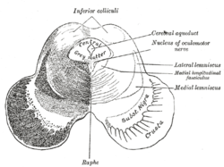
 Transverse section of mid-brain at level of inferior colliculi. (Medial longitudinal fasciculus labeled at center right.) | |
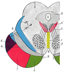
 Axial section through mid-brain. 1. Corpora quadrigemina. 2. Cerebral aqueduct. 3. Central gray stratum. 4. Interpeduncular space. 5. Sulcus lateralis. 6. Substantia nigra. 7. Red nucleus of tegmentum. 8. Oculomotor nerve, with 8’, its nucleus of origin. a. Lemniscus (in blue) with a’ the medial lemniscus and a" the lateral lemniscus. b. Medial longitudinal fasciculus. c. Raphe. d. Temporopontine fibers. e. Portion of medial lemniscus, which runs to the lentiform nucleus and insula. f. Cerebrospinal fibers. g. Frontopontine fibers. | |
| Details | |
| Identifiers | |
| Latin | fasciculus longitudinalis medialis |
| NeuroNames | 1588, 784 |
| NeuroLex ID | nlx_144065 |
| TA98 | A14.1.04.113 A14.1.05.304 A14.1.06.209 |
| TA2 | 5867 |
| FMA | 83846 |
| Anatomical terms of neuroanatomy | |
The medial longitudinal fasciculus (MLF) is a prominent bundle of nerve fibres which pass within the ventral/anterior portion of periaqueductal gray of the mesencephalon (midbrain).[1] It contains the interstitial nucleus of Cajal, responsible for oculomotor control, head posture, and vertical eye movement.[2]
The MLF interconnects interneurons of each abducens nucleus with motor neurons of the contralateral oculomotor nucleus; thus, the MLF mediates coordination of horizontal (side to side) eye movements, ensuring the two eyes move in unison (thus also enabling saccadic eye movements). The MLF also contains fibers projecting from the vestibular nuclei to the oculomotor and trochlear nuclei as well as the interstitial nucleus of Cajal; these connections ensure that eye movements are coordinated with head movements (as sensed by the vestibular system).[1]
The medial longitudinal fasciculus is the main central connection for the oculomotor nerve, trochlear nerve, and abducens nerve. It carries information about the direction that the eyes should move. Lesions of the medial longitudinal fasciculus can cause nystagmus and diplopia, which may be associated with multiple sclerosis, a neoplasm, or a stroke.
- ^ a b Waitzman, David M.; Oliver, Douglas L. (2002). "Midbrain". Encyclopedia of the Human Brain. Academic Press. pp. 43–68. doi:10.1016/B0-12-227210-2/00208-9. ISBN 978-0-12-227210-3.
- ^ Yu, Megan; Wang, Shu-Min (2022), "Neuroanatomy, Interstitial Nucleus of Cajal", StatPearls, Treasure Island (FL): StatPearls Publishing, PMID 31613454, retrieved 2022-03-12