| Mitochondrial myopathy | |
|---|---|
| Other names | Mitochondrial muscle disease; muscle mitochondrinopathy; muscle mitochondrial dysfunction |
 | |
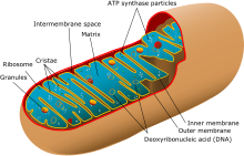
| Simplified structure of a typical mitochondrion | |
| Specialty | Neuromuscular medicine |
Mitochondrial myopathies are types of myopathies associated with mitochondrial disease.[1] Adenosine triphosphate (ATP), the chemical used to provide energy for the cell, cannot be produced sufficiently by oxidative phosphorylation when the mitochondrion is either damaged or missing necessary enzymes or transport proteins. With ATP production deficient in mitochondria, there is an over-reliance on anaerobic glycolysis which leads to lactic acidosis either at rest or exercise-induced.[2]
Primary mitochondrial myopathies are inherited, while secondary mitochondrial myopathies may be inherited (e.g. Duchenne's muscular dystrophy)[3] or environmental (e.g. alcoholic myopathy[4][5]). When it is an inherited primary disease, it is one of the metabolic myopathies.[6][4]
On biopsy, the muscle tissue of patients with these diseases usually demonstrate "ragged red" muscle fibers on Gomori trichrome staining. The ragged-red appearance is due to a buildup of abnormal mitochondria underneath the plasma membrane.[7] These ragged-red fibres may contain normal or abnormally increased accumulations of glycogen and neutral lipids, with histochemical staining showing abnormal respiratory chain involvement, such as decreased succinate dehydrogenase or cytochrome c oxidase.[8] Inheritance was believed to be maternal (non-Mendelian extranuclear). It is now known that certain nuclear DNA deletions can also cause mitochondrial myopathy such as the OPA1 gene deletion.[6]
- ^ "Mitochondrial Myopathy Information Page | National Institute of Neurological Disorders and Stroke". www.ninds.nih.gov. Retrieved 2017-02-28.
- ^ Cite error: The named reference
:5was invoked but never defined (see the help page). - ^ Cite error: The named reference
:9was invoked but never defined (see the help page). - ^ a b Simon L, Jolley SE, Molina PE (2017). "Alcoholic Myopathy: Pathophysiologic Mechanisms and Clinical Implications". Alcohol Research. 38 (2): 207–217. PMC 5513686. PMID 28988574.
- ^ Song BJ, Akbar M, Abdelmegeed MA, Byun K, Lee B, Yoon SK, Hardwick JP (2014-01-01). "Mitochondrial dysfunction and tissue injury by alcohol, high fat, nonalcoholic substances and pathological conditions through post-translational protein modifications". Redox Biology. 3: 109–123. doi:10.1016/j.redox.2014.10.004. PMC 4297931. PMID 25465468. S2CID 17113550.
- ^ a b Urtizberea JA, Severa G, Malfatti E (April 2023). "Metabolic Myopathies in the Era of Next-Generation Sequencing". Genes. 14 (5): 954. doi:10.3390/genes14050954. PMC 10217901. PMID 37239314.
- ^ "Ragged-red muscle fibers - MedGen - NCBI". www.ncbi.nlm.nih.gov. Retrieved 2024-01-05.
- ^ Sarnat, Harvey B.; Marín-García, José (May 2005). "Pathology of Mitochondrial Encephalomyopathies". Canadian Journal of Neurological Sciences. 32 (2): 152–166. doi:10.1017/S0317167100003929. ISSN 0317-1671. PMID 16018150. S2CID 1922603.