| Scintimammography | |
|---|---|
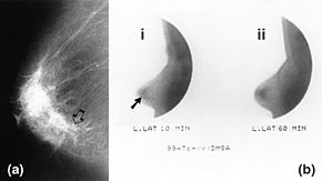
 Mammography (left) and DMSA scintimammography (right) images of 4.5cm breast carcinoma | |
| Synonyms | Nuclear medicine breast imaging Breast specific gamma imaging Breast scintigraphy Molecular breast imaging |
| ICD-10-PCS | CH1 |
| ICD-9-CM | 92.19 |
| HCPCS-L2 | S8080 |
Molecular breast imaging (MBI), also known as scintimammography, is a type of breast imaging test that is used to detect cancer cells in breast tissue of individuals who have had abnormal mammograms, especially for those who have dense breast tissue, post-operative scar tissue or breast implants.[1]
MBI is not used for screening or in place of a mammogram. Rather, it is used when the detection of breast abnormalities is not possible or not reliable on the basis of mammography and ultrasound alone. When mammography plus ultrasound are insufficient to characterize an abnormality, the gold standard next step is Magnetic Resonance Imaging (MRI) of the breast. However, in patients with contraindications (e.g. certain implantable devices) or who prefer to avoid MRI (claustrophobia, discomfort), use of scintimammography is an acceptable alternative.[2][1]
- ^ a b Muzahir, Saima (December 2020). "Molecular Breast Cancer Imaging in the Era of Precision Medicine". American Journal of Roentgenology. 215 (6): 1512–1519. doi:10.2214/AJR.20.22883. ISSN 0361-803X. PMID 33084364. S2CID 224823965.
- ^ Monticciolo, Debra L.; Newell, Mary S.; Moy, Linda; Niell, Bethany; Monsees, Barbara; Sickles, Edward A. (March 2018). "Breast Cancer Screening in Women at Higher-Than-Average Risk: Recommendations From the ACR". Journal of the American College of Radiology. 15 (3): 408–414. doi:10.1016/j.jacr.2017.11.034. ISSN 1546-1440. PMID 29371086.