| Mönckeberg's sclerosis | |
|---|---|
| Other names | Medial calcific sclerosis or Mönckeberg medial sclerosis |
 | |
| Microphotography of arterial wall with calcified (violet colour) atherosclerotic plaque (haematoxylin & eosin stain) | |
| Specialty | Cardiology |

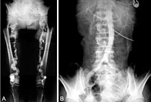
Mönckeberg's arteriosclerosis, or Mönckeberg's sclerosis, is a non-inflammatory form of arteriosclerosis (artery hardening), which differs from atherosclerosis traditionally. Calcium deposits are found in the muscular middle layer of the walls of arteries (the tunica media)[1] with no obstruction of the lumen. [2] It is an example of dystrophic calcification. This condition occurs as an age-related degenerative process. However, it can occur in pseudoxanthoma elasticum and idiopathic arterial calcification of infancy as a pathological condition, as well. Its clinical significance and cause are not well understood and its relationship to atherosclerosis and other forms of vascular calcification are the subject of disagreement.[3] Mönckeberg's arteriosclerosis is named after Johann Georg Mönckeberg,[4][5] who first described it in 1903.
The severity of calcium deposits formed by Mönckeberg's arteriosclerosis can be categorized into stages based on the histological appearance. Understanding these stages can help to understand disease progression and how the disease is caused. Stage 1 involves the formation of calcium deposits both inside and outside the vascular smooth muscle cells which compose the tunica media. Calcification outside of the vascular smooth muscle cells are commonly associated with damage to elastic fibers in the extra-cellular matrix. These calcium deposits also develop on the internal elastic lamina. Stage 2 and Stage 3 involve the formation of calcified sheaths spanning an increased diameter through the tunica media. Stage 4 then involves the formation of bony tissue from these calcifications through the process of osseous metaplasia.[6][7]
- ^ "Mönckeberg arteriosclerosis" at Dorland's Medical Dictionary
- ^ Fields C (June 2020). "Calciphylaxis and Mönckberg's Arteriosclerosis in a Diabetic Patient". Journal for Vascular Ultrasound. 44 (2): 83–88. doi:10.1177/1544316720923478. ISSN 1544-3167.
- ^ Micheletti RG, Fishbein GA, Currier JS, Singer EJ, Fishbein MC (August 2008). "Calcification of the internal elastic lamina of coronary arteries". Modern Pathology. 21 (8): 1019–1028. doi:10.1038/modpathol.2008.89. PMC 7029955. PMID 18536656.
- ^ synd/4064 at Who Named It?
- ^ J. G. Mönckeberg. Über die reine Mediaverkalkung der Extremitätenarterien und ihr Verhalten zur Arteriosklerose. Virchows Archiv für pathologische Anatomie und Physiologie, und für klinische Medicin, Berlin, 1903, 171: 141-167.
- ^ Lanzer P, Boehm M, Sorribas V, Thiriet M, Janzen J, Zeller T, et al. (June 2014). "Medial vascular calcification revisited: review and perspectives". European Heart Journal. 35 (23): 1515–1525. doi:10.1093/eurheartj/ehu163. PMC 4072893. PMID 24740885.
- ^ Razzaque MS (2013). "Phosphate toxicity and vascular mineralization". Contributions to Nephrology. 180: 74–85. doi:10.1159/000346784. ISBN 978-3-318-02369-5. PMID 23652551.