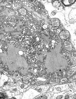

Negri bodies are eosinophilic, sharply outlined, pathognomonic inclusion bodies (2–10 μm in diameter) found in the cytoplasm of certain nerve cells containing the virus of rabies, especially in pyramidal cells[1] within Ammon's horn of the hippocampus. They are also often found in the Purkinje cells[1] of the cerebellar cortex from postmortem brain samples of rabies victims. They consist of ribonuclear proteins produced by the virus.[2]
They are named for Adelchi Negri.[3]
- ^ a b Sketchy Group, LLC. "2.3 Rhabdovirus". www.sketchymedical.com. Archived from the original on 2017-04-13. Retrieved 2017-04-12.
- ^ Lahaye, Xavier; Vidy, Aurore; Pomier, Carole; Obiang, Linda; Harper, Francis; Gaudin, Yves; Blondel, Danielle (15 August 2009). "Functional Characterization of Negri Bodies (NBs) in Rabies Virus-Infected Cells: Evidence that NBs Are Sites of Viral Transcription and Replication". Journal of Virology. 83 (16): 7948–7958. doi:10.1128/JVI.00554-09. ISSN 0022-538X. PMC 2715764. PMID 19494013.
- ^ synd/2491 at Who Named It?