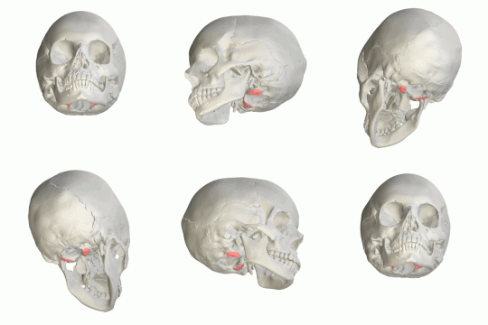
| Occipital condyles | |
|---|---|
 Occipital bone. Outer surface. Occipital condyles are shown in red. | |
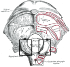
 Occipital bone. Outer surface. (Condyle for artic. with atlas labeled at lower left.) | |
| Details | |
| Identifiers | |
| Latin | condylus occipitalis |
| TA98 | A02.1.04.014 |
| TA2 | 557 |
| FMA | 52861 |
| Anatomical terminology | |
The occipital condyles are undersurface protuberances of the occipital bone in vertebrates, which function in articulation with the superior facets of the atlas vertebra.
The condyles are oval or reniform (kidney-shaped) in shape, and their anterior extremities, directed forward and medialward, are closer together than their posterior, and encroach on the basilar portion of the bone; the posterior extremities extend back to the level of the middle of the foramen magnum.
The articular surfaces of the condyles are convex from before backward and from side to side, and look downward and lateralward.
To their margins are attached the capsules of the atlanto-occipital joints, and on the medial side of each is a rough impression or tubercle for the alar ligament.
At the base of either condyle the bone is tunnelled by a short canal, the hypoglossal canal.