| Olfactory tubercle | |
|---|---|
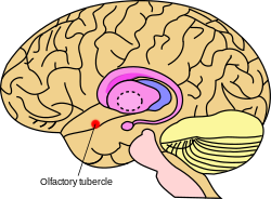
 Approximate location of the olfactory tubercle in the brain | |
| Details | |
| Part of | Mesolimbic pathway Ventral striatum; Olfactory cortex |
| Parts | Medial tubercle Lateral tubercle |
| Identifiers | |
| Latin | tuberculum olfactorium |
| Acronym(s) | OT |
| MeSH | D066208 |
| TA98 | A14.1.09.433 |
| TA2 | 5544 |
| Anatomical terminology | |
The olfactory tubercle (OT), also known as the tuberculum olfactorium, is a multi-sensory processing center that is contained within the olfactory cortex and ventral striatum and plays a role in reward cognition. The OT has also been shown to play a role in locomotor and attentional behaviors, particularly in relation to social and sensory responsiveness,[1] and it may be necessary for behavioral flexibility.[2] The OT is interconnected with numerous brain regions, especially the sensory, arousal, and reward centers, thus making it a potentially critical interface between processing of sensory information and the subsequent behavioral responses.[3]
The OT is a composite structure that receives direct input from the olfactory bulb and contains the morphological and histochemical characteristics of the ventral pallidum and the striatum of the forebrain.[4] The dopaminergic neurons of the mesolimbic pathway project onto the GABAergic medium spiny neurons of the nucleus accumbens and olfactory tubercle[5] (receptor D3 is abundant in these two areas [6]). In addition, the OT contains tightly packed cell clusters known as the islands of Calleja, which consist of granule cells. Even though it is part of the olfactory cortex and receives direct input from the olfactory bulb, it has not been shown to play a role in processing of odors.
- ^ Hitt, J. C.; Bryon, D. M.; Modianos, D. T. (1973). "Effects of rostral medial forebrain bundle and olfactory tubercle lesions upon sexual behavior of male rats". Journal of Comparative and Physiological Psychology. 82 (1): 30–36. doi:10.1037/h0033797. PMID 4567890.
- ^ Koob, G. F.; Riley, S. J.; Smith, S. C.; Robbins, T. W. (1978). "Effects of 6-hydroxydopamine lesions of the nucleus accumbens septi and olfactory tubercle on feeding, locomotor activity, and amphetamine anorexia in the rat". Journal of Comparative and Physiological Psychology. 92 (5): 917–927. doi:10.1037/h0077542. PMID 282297.
- ^ Wesson, D. W.; Wilson, D. A. (2011). "Sniffing out the contributions of the olfactory tubercle to the sense of smell: hedonics, sensory integration, and more?". Neuroscience & Biobehavioral Reviews. 35 (3): 655–668. doi:10.1016/j.neubiorev.2010.08.004. PMC 3005978. PMID 20800615.
- ^ Heimer, L.; Wilson, R. D. (1975). "The subcortical projections of the allocortex: Similarities in the neural connections of the hippocampus, the piriform cortex and the neocortex". In Santini, Maurizio (ed.). Golgi Centennial Symposium Proceedings. New York: Raven. pp. 177–193. ISBN 978-0911216806.
- ^ Ikemoto S (2010). "Brain reward circuitry beyond the mesolimbic dopamine system: a neurobiological theory". Neuroscience & Biobehavioral Reviews. 35 (2): 129–50. doi:10.1016/j.neubiorev.2010.02.001. PMC 2894302. PMID 20149820.
Recent studies on intracranial self-administration of neurochemicals (drugs) found that rats learn to self-administer various drugs into the mesolimbic dopamine structures–the posterior ventral tegmental area, medial shell nucleus accumbens and medial olfactory tubercle. ... In the 1970s it was recognized that the olfactory tubercle contains a striatal component, which is filled with GABAergic medium spiny neurons receiving glutamatergic inputs form cortical regions and dopaminergic inputs from the VTA and projecting to the ventral pallidum just like the nucleus accumbens
Figure 3: The ventral striatum and self-administration of amphetamine - ^ Brunton, Laurence L.; Hilal-Dandan, Randa; Knollmann, Bjorn C. (2018). Goodman & Gilman's - The pharmacological basis of therapeutics. Mc Graw Hill Education. p. 329. ISBN 978-1-25-958473-2.