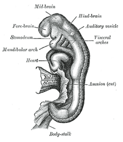
| Otic vesicle | |
|---|---|
 Embryo between eighteen and twenty-one days | |
 General formation of the otic vesicle | |
| Details | |
| Precursor | Otic placode, auditory pit or otic pit, otic cup, |
| Gives rise to | Membranous labyrinth of the inner ear |
| Identifiers | |
| Latin | vesicula otica |
| TE | vesicle_by_E5.15.1.0.0.0.4 E5.15.1.0.0.0.4 |
| Anatomical terminology | |
Otic vesicle, or auditory vesicle, consists of either of the two sac-like invaginations formed and subsequently closed off during embryonic development. It is part of the neural ectoderm, which will develop into the membranous labyrinth of the inner ear. This labyrinth is a continuous epithelium, giving rise to the vestibular system and auditory components of the inner ear.[1] During the earlier stages of embryogenesis, the otic placode invaginates to produce the otic cup. Thereafter, the otic cup closes off, creating the otic vesicle. Once formed, the otic vesicle will reside next to the neural tube medially, and on the lateral side will be paraxial mesoderm. Neural crest cells will migrate rostral and caudal to the placode.
The general sequence in formation of the otic vesicle is relatively conserved across vertebrates, although there is much variation in timing and stages.[2] Patterning during morphogenesis into the distinctive inner ear structures is determined by homeobox transcription factors including PAX2, DLX5 and DLX6, with the former specifying for ventral otic vesicle derived auditory structures and the latter two specifying for dorsal vestibular structures.[citation needed]
- ^ Freyer L, Aggarwal V, Morrow BE (December 2011). "Dual embryonic origin of the mammalian otic vesicle forming the inner ear". Development. 138 (24): 5403–14. doi:10.1242/dev.069849. PMC 3222214. PMID 22110056.
- ^ Park BY, Saint-Jeannet JP (December 2008). "Hindbrain-derived Wnt and Fgf signals cooperate to specify the otic placode in Xenopus". Developmental Biology. 324 (1): 108–21. doi:10.1016/j.ydbio.2008.09.009. PMC 2605947. PMID 18831968.