| Pallium | |
|---|---|
 | |
 | |
| Details | |
| Part of | Telencephalon |
| Identifiers | |
| Latin | pallium, or cortex cerebri |
| NeuroLex ID | birnlex_1494 |
| TA98 | A14.1.09.003 |
| TA2 | 5527 |
| TE | (neuroanatomy)_by_E5.14.3.4.3.1.30 E5.14.3.4.3.1.30 |
| Anatomical terms of neuroanatomy | |
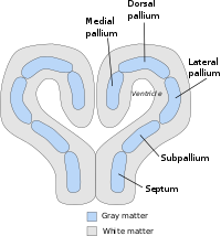
In neuroanatomy, pallium (pl.: pallia or palliums) refers to the layers of grey and white matter that cover the upper surface of the cerebrum in vertebrates. The non-pallial part of the telencephalon builds the subpallium. In basal vertebrates, the pallium is a relatively simple three-layered structure, encompassing 3–4 histogenetically distinct domains, plus the olfactory bulb.
It used to be thought that pallium equals cortex and subpallium equals telencephalic nuclei, but it has turned out, according to comparative evidence provided by molecular markers, that the pallium develops both cortical structures (allocortex and isocortex) and pallial nuclei (claustroamygdaloid complex), whereas the subpallium develops striatal, pallidal, diagonal-innominate and preoptic nuclei, plus the corticoid structure of the olfactory tuberculum.[1]
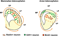
In mammals, the cortical part of the pallium registers a definite evolutionary step-up in complexity, forming the cerebral cortex, most of which consists of a progressively expanded six-layered portion isocortex, with simpler three-layered cortical regions allocortex at the margins. The allocortex subdivides into hippocampal allocortex, medially, and olfactory allocortex, laterally (including rostrally the olfactory bulb and anterior olfactory areas).
- ^ Fisher, Robin; Xie, Yuan-Yun (4 October 2010). "Growth Defects in the Dorsal Pallium after Genetically Targeted Ablation of Principal Preplate Neurons and Neuroblasts: A Morphometric Analysis". ASN Neuro. 2 (5): AN20100022. doi:10.1042/AN20100022. PMC 2949088. PMID 20957077.