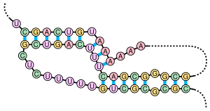

A pseudoknot is a nucleic acid secondary structure containing at least two stem-loop structures in which half of one stem is intercalated between the two halves of another stem. The pseudoknot was first recognized in the turnip yellow mosaic virus in 1982.[2] Pseudoknots fold into knot-shaped three-dimensional conformations but are not true topological knots. These structures are categorized as cross (X) topology within the circuit topology framework, which, in contrast to knot theory, is a contact-based approach.
- ^ Chen, JL; Greider, CW (7 June 2005). "Functional analysis of the pseudoknot structure in human telomerase RNA". Proceedings of the National Academy of Sciences of the United States of America. 102 (23): 8077–9. Bibcode:2005PNAS..102.8080C. doi:10.1073/pnas.0502259102. PMC 1149427. PMID 15849264.
- ^ Staple DW, Butcher SE (June 2005). "Pseudoknots: RNA structures with diverse functions". PLOS Biol. 3 (6): e213. doi:10.1371/journal.pbio.0030213. PMC 1149493. PMID 15941360.