| Pudendal canal | |
|---|---|
 | |
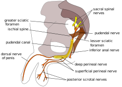
 Pudendal nerve and its course through the pudendal canal (labelled in yellow) | |
| Details | |
| Identifiers | |
| Latin | canalis pudendalis |
| TA98 | A09.5.04.003 |
| TA2 | 2436 |
| FMA | 22071 |
| Anatomical terminology | |
The pudendal canal (also called Alcock's canal) is an anatomical structure formed by the obturator fascia (fascia of the obturator internus muscle) lining the lateral wall of the ischioanal fossa. The internal pudendal artery and veins, and pudendal nerve pass through the pudendal canal, and the perineal nerve arises within it.[1]
- ^ "canalis pudendalis". TheFreeDictionary.com. Retrieved 2023-06-14.