This article needs more reliable medical references for verification or relies too heavily on primary sources. (May 2019) |  |
| Pulmonary consolidation | |
|---|---|
 | |
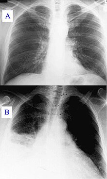
| Pneumonia as seen on chest X-ray. A: Normal chest X-ray. B: Abnormal chest X-ray with consolidation from pneumonia in the right lung, middle or inferior lobe (white area, left side of image). | |
| Specialty | Pulmonology |
A pulmonary consolidation is a region of normally compressible lung tissue that has filled with liquid instead of air.[1] The condition is marked by induration[2] (swelling or hardening of normally soft tissue) of a normally aerated lung. It is considered a radiologic sign. Consolidation occurs through accumulation of inflammatory cellular exudate in the alveoli and adjoining ducts. The liquid can be pulmonary edema, inflammatory exudate, pus, inhaled water, or blood (from bronchial tree or hemorrhage from a pulmonary artery). Consolidation must be present to diagnose pneumonia: the signs of lobar pneumonia are characteristic and clinically referred to as consolidation.[3]
- ^ "Consolidation – Definition". Merriam-Webster. Retrieved 2009-01-16.
- ^ "Induration- Definition". Merriam-Webster. Retrieved 2009-01-16.
- ^ Cite error: The named reference
Metlaywas invoked but never defined (see the help page).