| Rectus femoris muscle | |
|---|---|
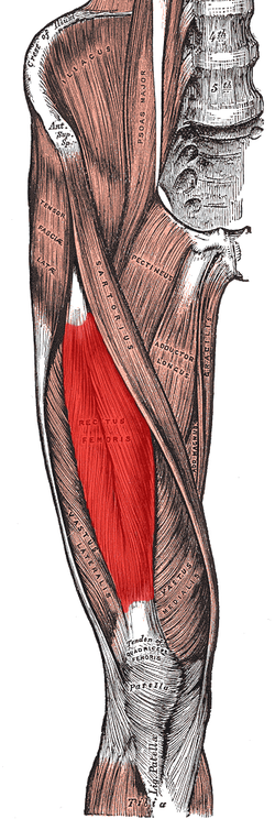
 Muscles of the iliac and anterior femoral regions. (Rectus femoris visible near center.) | |
| Details | |
| Pronunciation | /ˈrɛktəs ˈfɛmərɪs/ |
| Origin | Anterior inferior iliac spine and the exterior surface of the bony ridge which forms the groove on the iliac portion of the acetabulum |
| Insertion | Inserts into the patellar tendon as one of the four quadriceps muscles |
| Artery | Descending branch of the lateral femoral circumflex artery |
| Nerve | Femoral nerve |
| Actions | Knee extension; hip flexion |
| Antagonist | Hamstring |
| Identifiers | |
| Latin | musculus rectus femoris |
| TA98 | A04.7.02.018 |
| TA2 | 2614 |
| FMA | 22430 |
| Anatomical terms of muscle | |
The rectus femoris muscle is one of the four quadriceps muscles of the human body. The others are the vastus medialis, the vastus intermedius (deep to the rectus femoris), and the vastus lateralis. All four parts of the quadriceps muscle attach to the patella (knee cap) by the quadriceps tendon.
The rectus femoris is situated in the middle of the front of the thigh; it is fusiform in shape, and its superficial fibers are arranged in a bipenniform manner, the deep fibers running straight (Latin: rectus) down to the deep aponeurosis. Its functions are to flex the thigh at the hip joint and to extend the leg at the knee joint.[1]
- ^ Elizabeth Quinn. "Function of the Rectus Femoris Muscle". Verywell Fit. Retrieved 2022-07-23.