| Renal pelvis | |
|---|---|
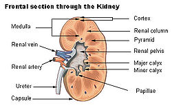
 Cross-section of the kidney, with major structures labelled. The renal pelvis, located in the middle of the image, collects urine from the urinary calices. | |
 An image showing just the pelvis and calices of the kidneys, with the rest of the kidney removed, from a dissected cow and seal specimen. These vary greatly in size and number depending on species.[citation needed] | |
| Details | |
| Precursor | Ureteric bud |
| System | Urinary system |
| Identifiers | |
| Latin | pelvis renalis |
| MeSH | D007682 |
| TA98 | A08.1.05.001 |
| TA2 | 3384 |
| FMA | 15575 |
| Anatomical terminology | |
The renal pelvis or pelvis of the kidney is the funnel-like dilated part of the ureter in the kidney. It is formed by the convergence of the major calyces, acting as a funnel for urine flowing from the major calyces to the ureter. It has a mucous membrane and is covered with transitional epithelium and an underlying lamina propria of loose-to-dense connective tissue.
The renal pelvis is situated within the renal sinus alongside the other structures of the renal sinus.[1]
- ^ Sobotta anatomy textbook. Friedrich Paulsen, Tobias M. Böckers, J. Waschke, Stephan Winkler, Katja Dalkowski, Jörg Mair, Sonja Klebe, Elsevier ClinicalKey. Munich. 2019. p. 354. ISBN 978-0-7020-6760-0. OCLC 1132300315.
{{cite book}}: CS1 maint: location missing publisher (link) CS1 maint: others (link)