| |||

| |||
| |||
| Names | |||
|---|---|---|---|
| Preferred IUPAC name
3,7-Diamino-2,8-dimethyl-5-phenylphenazin-5-ium chloride | |||
| Identifiers | |||
3D model (JSmol)
|
|||
| ChEBI | |||
| ChemSpider | |||
| ECHA InfoCard | 100.006.836 | ||
PubChem CID
|
|||
| UNII | |||
CompTox Dashboard (EPA)
|
|||
| |||
| |||
| Properties | |||
| C20H19ClN4 | |||
| Molar mass | 350.85 g·mol−1 | ||
| Soluble | |||
| Hazards | |||
| GHS labelling: | |||
  [1] [1]
| |||
| Danger[1] | |||
| H315, H318[1] | |||
| P264, P280, P302+P352, P305+P351+P338, P310, P332+P313, P362[1] | |||
| NFPA 704 (fire diamond) | |||
Except where otherwise noted, data are given for materials in their standard state (at 25 °C [77 °F], 100 kPa).
| |||
Safranin (Safranin O or basic red 2) is a biological stain used in histology and cytology. Safranin is used as a counterstain in some staining protocols, colouring cell nuclei red. This is the classic counterstain in both Gram stains and endospore staining. It can also be used for the detection of cartilage,[2] mucin and mast cell granules.
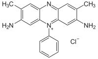
Safranin typically has the chemical structure shown at right (sometimes described as dimethyl safranin). There is also trimethyl safranin, which has an added methyl group in the ortho- position (see Arene substitution pattern) of the lower ring. Both compounds behave essentially identically in biological staining applications, and most manufacturers of safranin do not distinguish between the two. Commercial safranin preparations often contain a blend of both types.
Safranin is also used as redox indicator in analytical chemistry.
- ^ a b c d "Safety Data Sheet: Safranin O" (PDF). LabChem. Archived from the original (PDF) on 10 March 2016. Retrieved 10 March 2016.
- ^ Rosenberg L (1971). "Chemical Basis for the Histological Use of Safranin O in the Study of Articular Cartilage". J Bone Joint Surg Am. 53 (1): 69–82. doi:10.2106/00004623-197153010-00007. PMID 4250366. Archived from the original (abstract) on 2008-04-17.


