This article needs additional citations for verification. (December 2008) |
| Trabecular meshwork | |
|---|---|
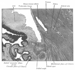
 Enlarged general view of the iridial angle. (When enlarged, visible with older label of 'trabecular tissue') | |
| Details | |
| Identifiers | |
| Latin | reticulum trabeculare sclerae |
| MeSH | D014129 |
| Anatomical terminology | |


The trabecular meshwork is an area of tissue in the eye located around the base of the cornea, near the ciliary body, and is responsible for draining the aqueous humor from the eye via the anterior chamber (the chamber on the front of the eye covered by the cornea).
The tissue is spongy and lined by trabeculocytes; it allows fluid to drain into a set of tubes called Schlemm's canal which is lined by endothelium with blood and lymphatic properties that allow aqueous humor to flow into the blood system.[1]
- ^ Karpinich NO, Caron KM (September 2014). "Schlemm's canal: more than meets the eye, lymphatics in disguise". The Journal of Clinical Investigation. 124 (9): 3701–3. doi:10.1172/JCI77507. PMC 4151199. PMID 25061871.